. 2009 Nov 12;114(20):4441-50.
doi: 10.1182/blood-2009-07-233718. Epub 2009 Sep 17.

Diverse marrow stromal cells protect CLL cells from spontaneous and drug-induced apoptosis: development of a reliable and reproducible system to assess stromal cell adhesion-mediated drug resistance

Affiliations

Diverse marrow stromal cells protect CLL cells from spontaneous and drug-induced apoptosis: development of a reliable and reproducible system to assess stromal cell adhesion-mediated drug resistance

Antonina V Kurtova et al. Blood. .

Abstract

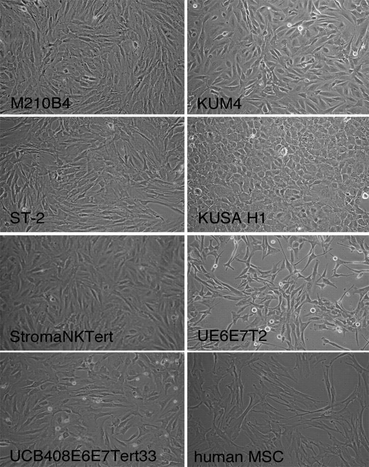
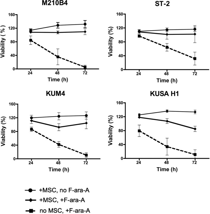
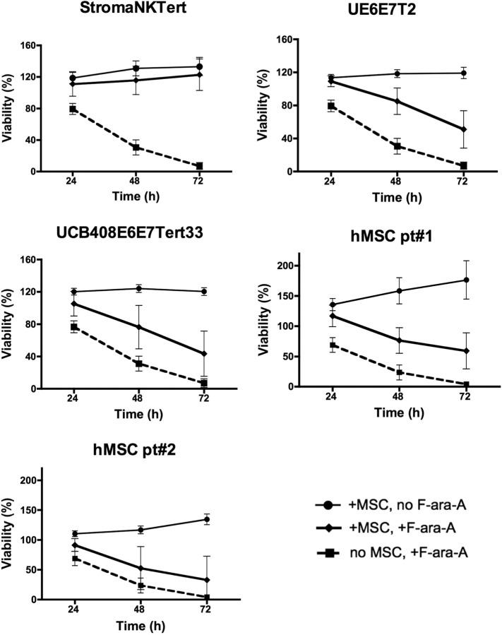
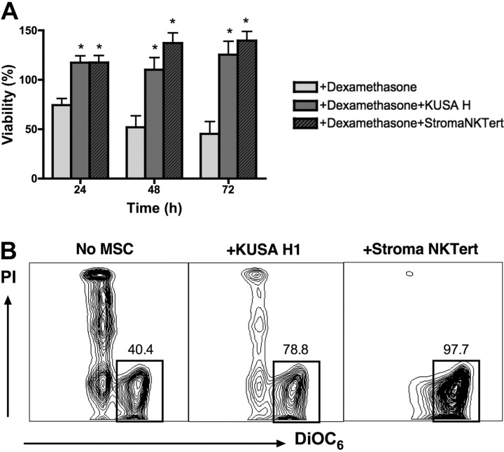
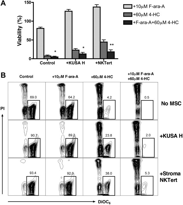
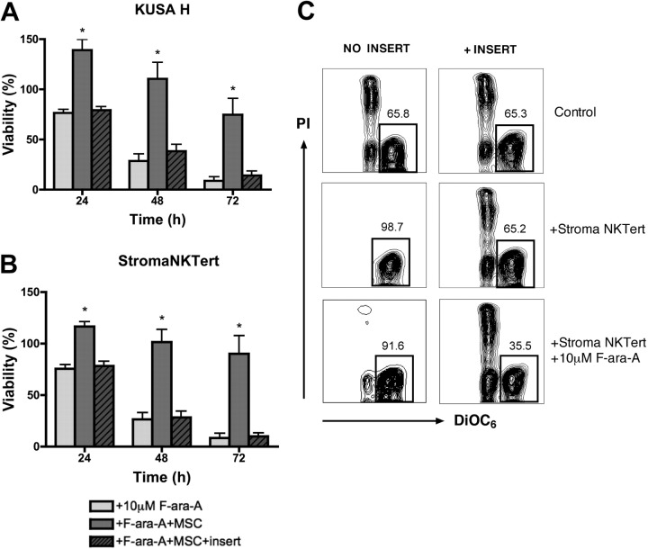
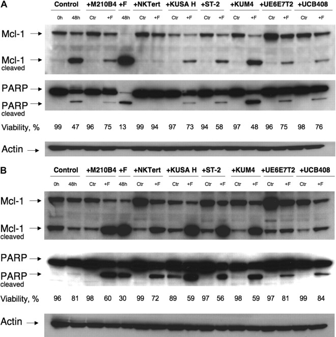
Marrow stromal cells (MSCs) provide important survival and drug resistance signals to chronic lymphocytic leukemia (CLL) cells, but current models to analyze CLL-MSC interactions are heterogeneous. Therefore, we tested different human and murine MSC lines and primary human MSCs for their ability to protect CLL cells from spontaneous and drug-induced apoptosis. Our results show that both human and murine MSCs are equally effective in protecting CLL cells from fludarabine-induced apoptosis. This protective effect was sustained over a wide range of CLL-MSC ratios (5:1 to 100:1), and the levels of protection were reproducible in 4 different laboratories. Human and murine MSCs also protected CLL cells from dexamethasone- and cyclophosphamide-induced apoptosis. This protection required cell-cell contact and was virtually absent when CLL cells were separated from the MSCs by micropore filters. Furthermore, MSCs maintained Mcl-1 and protected CLL cells from spontaneous and fludarabine-induced Mcl-1 and PARP cleavage. Collectively, these studies define common denominators for CLL cocultures with MSCs. They also provide a reliable, validated tool for future investigations into the mechanism of MSC-CLL cross talk and for drug testing in a more relevant fashion than the commonly used suspension cultures.


Figures

Figure 1
Figure 1
Phenotype of the different MSCs. Figure displays phase-contrast photomicrographs that depict the morphologic appearance of MSC lines and primary MSCs derived from the marrow of a patient with CLL. Cells were imaged in medium using a phase-contrast microscope (Model ELWD 0.3; Nikon) with a 10 × /0.25 NA objective lens. Images were captured with a Nikon D40 digital camera (Nikon Corp) with the use of Camera Control Pro software (Nikon); when necessary, Adobe Photoshop 9.0 (Adobe Systems) was used for image processing.
Figure 2
Figure 2
Murine MSCs protect CLL cells from spontaneous and F-ara-A–induced apoptosis. Displayed are the mean relative viabilities of CLL cells in absence or presence of murine MSCs and/or 10 μM F-ara-A at the time points shown on the horizontal axis. Results represent data for 20:1 CLL/MSC ratios and are the mean (± SEM) relative viabilities, compared with untreated controls (100%) from 5 different CLL patients for each MSC cell line. All murine MSC lines provided significant levels of protection from F-ara-A–induced apoptosis. The names of the murine MSCs are displayed above each diagram.
Figure 3
Figure 3
Human MSCs protect CLL cells from spontaneous and drug-induced apoptosis. Displayed are the mean relative viabilities of CLL cells in the absence or presence of murine MSCs and/or 10 μM F-ara-A at the time points shown on the horizontal axis. Results represent data for 20:1 CLL/MSC ratios and are the mean (± SEM) relative viabilities, compared with untreated controls (100%) from 3 to 5 different patients with CLL for each MSC line. All human MSC lines (except for the primary hMSC pt#2; see supplemental Table 3) provided significant levels of protection from F-ara-A–induced apoptosis, although the levels of protection generally were lower than those provided by murine MSCs. The names of the human MSCs are displayed above each diagram.
Figure 4
Figure 4
Coculture with MSCs protects CLL cells from dexamethasone-induced apoptosis. (A) Bar diagram depicts the mean (± SEM) relative viabilities of CLL cells treated with dexamethasone in the presence or absence of murine (KUSA H) or human (StromaNKTert) MSCs at the time points displayed on the horizontal axis from 5 different patients with CLL. *Significant protection from dexamethasone-induced cytotoxicity compared with control sample (P < .05). (B) Contour plots from a representative CLL sample depict viability of CLL cells, as determined by staining with DiOC6 and PI, after 48 hours of incubation with 10 μM dexamethasone in absence or presence of MSCs, as indicated above each of the plots. The percentage of viable cells is displayed above each of the gates that define viable cells (DiOC6bright PIexclusion).
Figure 5
Figure 5
4-HC and combinations of 4-HC and F-ara-A induce high levels of cytotoxicity, even in the presence of MSCs. CLL cells were cultured with or without MSCs and with 10 μM F-ara-A and/or 60 μM 4-HC. (A) Bar diagram depicts the mean (± SEM) relative viabilities of CLL cells treated with F-ara-A, 4-HC, or a combination of F-ara-A and 4-HC after 24 hours. Results are presented as mean relative viability compared with untreated controls (100%) and are the mean (± SEM) viabilities of CLL samples from 8 different patients. * and ** indicate significant increases in cytotoxicity of the combination of 4-HC plus F-ara-A compared with 4-HC alone (P < .05 or P < .01). (B) Contour plots show the viability of CLL cells after 24 hours of incubation for 1 representative patient. The percentage of viable cells (DiOC6bright PIexclusion) is shown above each of the gates.
Figure 6
Figure 6
Direct cell-to-cell contact is essential for cell adhesion–mediated drug resistance. CLL cells were treated with 10 μM F-ara-A and incubated with KUSA H1 (A) and StromaNKTert (B) in the presence or absence of micropore membrane insert for 24, 48, and 72 hours. Bars represent the mean viability of CLL cells compared with untreated control (100%). Data shown are the mean (± SEM) of 6 independent experiments. *Significant protection of CLL cells from F-ara-A–induced apoptosis in direct CLL–MSC contact compared with control sample (P < .05). (C) Presented are contour plots of CLL cells from a representative patient after 48 hours of coculture with StromaNKTert in the conditions indicated above and on the side of the plots. The relative percentages of viable cells are displayed above each of the gates. Direct cell-to-cell contact is essential not only for protection from fludarabine-induced apoptosis but also from spontaneous apoptosis.
Figure 7
Figure 7
Mcl-1 and PARP expression in CLL cells cocultured with MSCs. CLL cells were cultured with the MSC lines displayed on the top horizontal axes or in suspension (“Control”) for 48 hours with or without 10 μM F-ara-A (labeled “Ctr” or “+F,” respectively). Then, cleaved and uncleaved Mcl-1 and PARP were analyzed by Western blotting, and the respective immunobands are indicated on the left-hand side. Cell viability for each condition was measured by flow cytometry, and the percentage of viable cells is displayed below each of the blots. In most cases, MSCs coculture up-regulated Mcl-1 and PARP expression compared with CLL cells in suspension (control at 48 hours vs the “Ctr” bands in the presence of MSCs). Suspension culture of CLL cells results in spontaneous apoptosis, with associated Mcl-1 and PARP cleavage (panels A-B 2nd lane from the left), which was paralleled by a decrease in CLL cell viability from 99% to 47% in panel A and from 96% to 81% in panel B. Treatment with fludarabine resulted in cleavage of the majority of Mcl-1 and PARP in the absence of MSCs (panels A-B 5th lane from the left). This was largely inhibited, sometimes almost abrogated (eg, panel A lanes 6-7) by the presence of MSCs. Displayed are Western blots of CLL B-cell lysates from 2 representative patients (A-B).

References

    1. Tam CS, O'Brien S, Wierda W, et al. Long-term results of the fludarabine, cyclophosphamide, and rituximab regimen as initial therapy of chronic lymphocytic leukemia. Blood. 2008;112(4):975–980. - PMC - PubMed
    1. Rawstron AC, Kennedy B, Evans PA, et al. Quantitation of minimal disease levels in chronic lymphocytic leukemia using a sensitive flow cytometric assay improves the prediction of outcome and can be used to optimize therapy. Blood. 2001;98(1):29–35. - PubMed
    1. Lagneaux L, Delforge A, Bron D, De Bruyn C, Stryckmans P. Chronic lymphocytic leukemic B cells but not normal B cells are rescued from apoptosis by contact with normal bone marrow stromal cells. Blood. 1998;91(7):2387–2396. - PubMed
    1. Damiano JS, Cress AE, Hazlehurst LA, Shtil AA, Dalton WS. Cell adhesion mediated drug resistance (CAM-DR): role of integrins and resistance to apoptosis in human myeloma cell lines. Blood. 1999;93(5):1658–1667. - PMC - PubMed
    1. Burger JA, Kipps TJ. Chemokine receptors and stromal cells in the homing and homeostasis of chronic lymphocytic leukemia B cells. Leuk Lymphoma. 2002;43(3):461–466. - PubMed

Publication types

MeSH terms

Substances