. 2009 Nov;20(11):2315-27.
doi: 10.1681/ASN.2008111203. Epub 2009 Sep 17.

Proliferation and migration of label-retaining cells of the kidney papilla

Affiliations

Proliferation and migration of label-retaining cells of the kidney papilla

Juan A Oliver et al. J Am Soc Nephrol. 2009 Nov.

Abstract

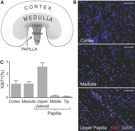
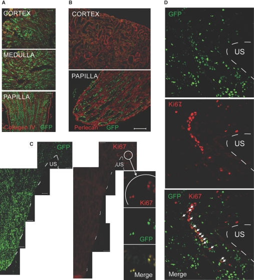
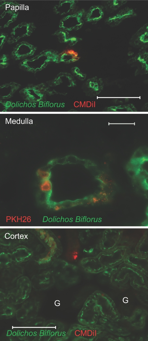
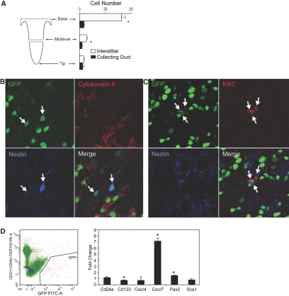
The kidney papilla contains a population of cells with several characteristics of adult stem cells, including the retention of proliferation markers during long chase periods (i.e., they are label-retaining cells [LRCs]). To determine whether the papillary LRCs generate new cells in the normal adult kidney, we examined cell proliferation throughout the kidney and found that the upper papilla is a site of enhanced cell cycling. Using genetically modified mice that conditionally expressed green fluorescence protein fused to histone 2B, we observed that the LRCs of the papilla proliferated only in its upper part, where they associate with "chains" of cycling cells. The papillary LRCs decreased in number with age, suggesting that the cells migrated to the upper papilla before entering the cell cycle. To test this directly, we marked papillary cells with vital dyes in vivo and found that some cells in the kidney papilla, including LRCs, migrated toward other parts of the kidney. Acute kidney injury enhanced both cell migration and proliferation. These results suggest that during normal homeostasis, LRCs of the kidney papilla (or their immediate progeny) migrate to the upper papilla and form a compartment of rapidly proliferating cells, which may play a role in repair after ischemic injury.


Figures

Figure 1.
Figure 1.
Cellular proliferation in adult kidney is shown. (A) Kidney regions were examined for cellular proliferation. (B) Ki67-positive cells in representative sections of kidney cortex, medulla, and upper papilla are shown. Cell nuclei stained with DAPI (blue) and Ki67 stained with rhodamine (red) are shown. Bars = 50 μm. (C) Fraction of Ki67-positive cells is shown. Cortex and medulla had a similar number of Ki67-positive cells, but the lateral side of the upper papilla had significantly more Ki67-positive cells than both the cortex and the medulla as well as other parts of the papilla (P < 0.01). The tip and middle part of the papilla had significantly fewer Ki67-positive cells than all other areas of the kidney (P < 0.01).
Figure 2.
Figure 2.
Papillary LRCs in mice expressing histone 2B-GFP are shown. (A) Cortex, medulla, and papilla from a 10-d-old mouse pulsed with doxycycline during embryonic life are shown. (B) Low-cycling cells in kidney papilla are shown. A mouse was pulsed with doxycycline during embryonic life and chased for 11 wk after birth; shown are kidney cortex and papilla. Papillary LRCs retain GFP. Extracellular matrix labeled with an antibody to perlecan is shown. Bar = 50 μm. (C) Papillary LRCs proliferate in the upper papilla. Shown is kidney papilla with multiple LRCs; broken white line depicts papillary edge. Proliferating cells (Ki67 positive) were restricted to the upper papilla, and high-magnification views of the Ki67-positive cells showed them also to be GFP+ (i.e., low-cycling cells). Bars = 50 μm. (D) Chains of proliferating cells in upper papilla contain LRCs: Papillary LRCs (GFP+, top) and proliferating cells (Ki67-positive, middle). The merged picture shows that several cells are positive for both Ki67 and GFP (arrows). Broken while line depicts papillary edge. US, urinary space.
Figure 3.
Figure 3.
Papillary LRCs decrease with age. (A) Shown is the frequency of BrdU-retaining cells in papillae of 3- and 12-mo-old rats (n = 4 for each age). There was a marked decrease in the number of BrdU-retaining cells in the 12-mo-old rats (P < 0.001). (B) Shown is the location of BrdU-retaining cells in papilla of a 3-mo-old rat. (C) Shown is the location of BrdU-retaining cells in papilla of a 12-mo-old rat. Cross-sectional views were taken of mid-papilla labeled with Dolichos biflorus agglutinin-Rhodamine (identifies collecting ducts) and BrdU-antibody (FITC) of papillae from 3- and 12-mo-old rats. In the 3-mo-old rat, BrdU-retaining cells were abundant at both the center and the periphery of the papilla, whereas in the 12-mo-old rat, few BrdU-retaining cells remained at the center of the papilla. US, urinary space.
Figure 4.
Figure 4.
In vivo labeling and migration of renal papillary cells are shown. (A) Cells in the superficial papilla of rat were labeled. Rat kidney papilla were labeled with KPH26 and isolated 1 h later. Collecting ducts were labeled with Dolichos biflorus agglutinin-FITC (photomicrograph of 25-μm section). Bar = 200 μm. (B) Papillary cells labeled with dye show upward migration; these are representative sections of normal kidneys 3 d after introduction of dye into the urinary space. (Top) Upper papilla shows CMDiI-containing cells; collecting ducts were labeled with Dolichos biflorus agglutinin-FITC. Bar = 20 μm. (Bottom) Kidney medulla shows cells containing PKH26, and thick ascending limbs were labeled with antibody to Tamm-Horsfall protein–FITC (photomicrographs of 25-μm sections). Bar = 100 μm. (C) Cells in the superficial papilla of H2B-GFP mouse were labeled. Note GFP+ cells in the body of the papilla. (D) Papillary LRCs labeled with dye show upward migration. Note that some cells in the upper papilla (top) and medulla (bottom) that contain injected CMDiI (red) were also LRCs (had a GFP+ nucleus). Bars = 20 μm
Figure 5.
Figure 5.
Cellular proliferation occurred in upper papilla after ischemic injury. (A) There was a selective increase of cells in S phase in the rat upper papilla during the first hour after injury. Shown is the lateral part of the upper papilla from a rat subjected to 45 min of unilateral renal ischemia (left) and from a control normal rat (right) both given BrdU for 1 h. Note abundant BrdU-labeled cells in the postischemic papilla, whereas the cortex and medulla of the same kidney revealed very rare BrdU-labeled cells. US, urinary space. Photomicrographs were done in 100-μm tissue sections obtained with a vibratome. Bars = 100 μm. (B) Papillary LRCs in the upper papilla proliferate after ischemic injury. Rat upper papilla have BrdU-retaining cells 12 h after injury; Ki67-positive cells (i.e., proliferating) formed chain-like structures, and several of these cells contained BrdU, indicative of low-cycling history (n = 2). (C) There is a selective increase of proliferating cells in the upper papilla of an H2B-GFP mouse subjected to 20 min of unilateral renal ischemia. Twenty-four hours later, there were abundant Ki67-positive cells in the postischemic upper papilla, whereas cycling cells were very rare in the contralateral organ (right). Bars = 50 μm. (D) Some proliferating cells in the upper papilla of H2B-GFP mouse were LRCs. Confocal microphotograph with ortho projections show that several LRCs (GFP+) from the postischemia section in C also contained Ki67. (E) Decrease in LRCs occurs 2 wk after ischemia. LRCs (GFP+) are seen in the upper papillae from an H2B-GFP mouse subjected to unilateral transient ischemia. (Left) Papilla of the kidneys were subjected to ischemia. (Right) The contralateral, control side is shown. Bars = 50 μm.
Figure 6.
Figure 6.
Papillary cells labeled with dye after injury show upward migration. Labeled cells with dye were easily detected in the parenchyma of the papilla; collecting ducts were labeled with Dolichos biflorus agglutinin–FITC (top; bar = 100 μm). The kidney medulla also contained cells with dye, some of which integrated into collecting ducts (bar = 20 μm). Only in kidneys subjected to ischemic injury were cells with dye occasionally detected in the kidney cortex. G, glomerulus. Bar = 100 μm (photomicrographs of 25-μm sections).
Figure 7.
Figure 7.
(A) LRCs were identified in rat kidney papilla. Kidney papillae from 1-yr-old rats (n = 5) that had been pulse-chased with BrdU were isolated from the rest of the kidney and sectioned as shown (left). (Right) LRCs in the tip of the papilla were similarly located in the collecting duct and in the interstitial space, whereas in the middle level of the papilla, there was a slight but statistically significant greater number of LRC interstitial cells than collecting duct cells. This difference was markedly accentuated at the base of the papilla. (B) LRCs were identified in H2B-GFP mouse. Confocal image of a papillary tip of a H2B-GFP mouse show LRCs (GFP) in the collecting ducts (cytokeratin 8 positive; Rhodamine) and interstitium. Note in the merged image that two LRCs that are outside the collecting ducts (marked with arrows) are nestin positive (blue). (C) LRCs in H2B-GFP mouse proliferate. Confocal image of the upper part of the papilla shows three LRCs (arrows) that are cycling (Ki67 positive; Rhodamine) and nestin (blue) positive and hence are interstitial cells. When the same area was stained with cytokeratin 8 to identify LRCs that were also proliferating (Ki67 positive), none was found. (D) FACS isolation and relative overexpression of mRNA in GFP+ versus GFP cells are shown. Cells from kidney papilla that were negative for DAPI (i.e., alive cells) and PE-labeled antibodies to CD45, TER119-PE, and CD31 were sorted to GFP+ and GFP populations. Of the genes tested, Cd24a, Cd133, Cxcr7, and Pax2 were significantly overexpressed (fold change) in the papillary LRCs, whereas the expression of Cxcr4 and of Sca1 was no different between the two cell populations. Expression of Oct4 was not detected in either cell population. *P < 0.05.

Comment in

References

    1. Lavker RM, Sun TT: Epidermal stem cells: Properties, markers, and location. Proc Natl Acad Sci U S A 97: 13473–13475, 2000 - PMC - PubMed
    1. Beltrami AP, Barlucchi L, Torella D, Baker M, Limana F, Chimenti S, Kasahara H, Rota M, Musso E, Urbanek K, Leri A, Kajstura J, Nadal-Ginard B, Anversa P: Adult cardiac stem cells are multipotent and support myocardial regeneration. Cell 114: 658–659, 2003 - PubMed
    1. Petersen BE, Zajac VF, Michalopoulos GK: Hepatic oval cell activation in response to injury following chemically induced periportal or pericentral damage in rats. Hepatology 27: 1030–1038, 1998 - PubMed
    1. Mothe AJ, Tator CH: Proliferation, migration, and differentiation of endogenous ependymal region stem/progenitor cells following minimal spinal cord injury in the adult bat. Neuroscience 131: 177–187, 2005 - PubMed
    1. Taylor G, Lehrer MS, Jensen PJ, Sun TT, Lavker RM: Involvement of follicular stem cells in forming not only the follicle but also the epidermis. Cell 102: 451–461, 2000 - PubMed

Publication types