. 2009 Oct 1;23(19):2284-93.
doi: 10.1101/gad.1837009. Epub 2009 Sep 17.

An inducible change in Fox-1/A2BP1 splicing modulates the alternative splicing of downstream neuronal target exons

Affiliations

An inducible change in Fox-1/A2BP1 splicing modulates the alternative splicing of downstream neuronal target exons

Ji-Ann Lee et al. Genes Dev. .

Abstract

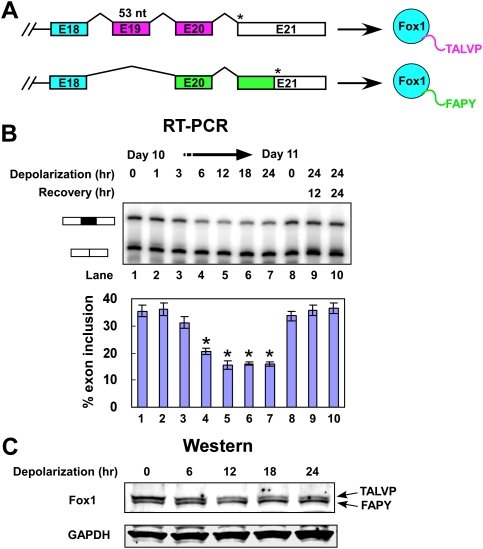
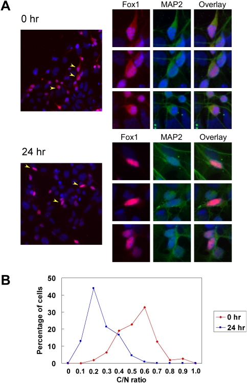
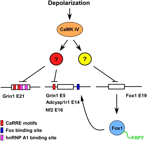
Neuronal depolarization and CaM kinase IV signaling alter the splicing of multiple exons in transcripts for ion channels, neurotransmitter receptors, and other synaptic proteins. These splicing changes are mediated in part by special CaM kinase-responsive RNA elements, within or adjacent to exons that are repressed in the initial phase of chronic depolarization. The splicing of many neuronal transcripts is also regulated by members of the Fox (Feminizing gene on X) protein family, and these Fox targets are also often proteins affecting synaptic activity. We show that Fox-1/Ataxin 2-Binding Protein 1 (A2BP1), a protein implicated in a variety of neurological diseases, can counteract the effects of chronic depolarization on splicing. We find that exon 19 of Fox-1 is itself repressed by depolarization. Fox-1 transcripts missing exon 19 encode a nuclear isoform of Fox-1 that progressively replaces the cytoplasmic Fox-1 isoform as cells are maintained depolarizing media. The resulting increase in nuclear Fox-1 leads to the reactivation of many Fox-1 target exons, including exon 5 of the NMDA receptor 1, that were initially repressed by the high-KCl medium. These results reveal a novel mechanism for the slow modulation of splicing as cells adapt to chronic stimuli: The subcellular localization of a splicing regulator is controlled through its own alternative splicing.


Figures

Figure 1.
Figure 1.
Depolarization represses splicing of Fox-1 E19 in differentiated P19 cells. (A) Diagram of the alternative splicing within the 3′ region of the Fox-1 pre-mRNA. Alternative splicing of E19 alters the reading frame to generate two C-terminal peptide sequences, depicted as purple and green bars in the pre-mRNA. Asterisks (*) indicate the stop codons in the transcripts. The C-terminal peptides are indicated to the right. (B) Denaturing gel electrophoresis of Fox-1 RT–PCR products. The E19-included and E19-excluded bands are indicated. (Lanes 1–7) P19 cells were differentiated for 10 d and then depolarized for 0, 1, 3, 6, 12, 18, 24 h. Also shown are RNA from day 11 untreated cells (lane 8), and from cells depolarized for 24 h followed by recovery for 12 and 24 h (lanes 9,10). A graph of E19 inclusion is shown below the gel. Error bars represent SD; n = 3. Asterisks denote significant changes in splicing, indicated by a t-test P-value of <0.05. (C) Immunoblot analysis of Fox-1 collected from the same cells as in B. The identities of the two Fox-1 isoforms are indicated, and GAPDH was used as loading control for total protein. The protein samples were separated on a 4%–20% gradient gel.
Figure 2.
Figure 2.
The subcellular distribution of Fox-1 protein is changed after depolarization. (A) Immunostaining of Fox-1 protein in differentiated P19 cells. Fox-1 (red) is found in the cytoplasm and nucleus in untreated cells (0 h, top left panel) and becomes mainly nuclear after depolarization (24 h of depolarization, bottom left panel). Three representative cells from each field (arrowheads) are shown at the right, with the individual channels of Fox-1 (red), cytoplasmic neuronal marker MAP2 (green), and the overlay indicated. DNA stain DAPI (blue) is shown in every panel. (B) The ratio of C/N Fox-1 was measured in 110 rested cells (0 h) and 132 depolarized cells (24 h). The C/N ratio significantly decreases after depolarization, with the peak ratio shifting from 0.6 in the rested cells to 0.2 in the depolarized cells (P-value = 2.82 × 10−41 from a two-sample t-test).
Figure 3.
Figure 3.
Fox-1-FAPY activates splicing of exons carrying downstream Fox-binding sites. (A) Immunoblot of Fox-2 and Fox-1 proteins in N2A cells. N2A cells were transfected with a plasmid expressing a shRNA targeting the Fox-2 3′ UTR (shFox2) or with a control vector (shV). Forty-eight hours after shRNA transfection, N2A cells were retransfected with Flag-Fox-1-FAPY (F1F), Flag-Fox-1-TALVP (F1T), or control (Vec) expression vectors; incubated for 24 h; and immunoblotted for Fox-2 and Fox-1 proteins. GAPDH served as a loading control. (B) RT–PCR analyses of exons carrying a Fox-binding site, UGCAUG (blue box) in the downstream intron (left panels), and control exons that lack Fox-binding sites (right panels). The exon IDs are shown above the gels. The cell conditions are as in A. Graphs of percent exon inclusion (% Ex In) are shown to the right of the gels. Error bars represent SD; n = 3.
Figure 4.
Figure 4.
Fox-1 antagonizes depolarization and CaMKIV-induced splicing repression. (A) The splicing repression of Fox-dependent exons is relieved during chronic depolarization. The inclusion levels of exons carrying a UGCAUG element within the first 200 nt of the downstream intron are shown in the left graph. The splicing of exons without Fox-binding sites are shown in the right graph. Exon inclusion was measured in untreated cells (0 h) and in cells treated with 50 mM KCl for 6, 12, 18, and 24 h. Error bars represent SD; n = 3. Representative RT–PCR gels for NR1 (Grin1) exon 5 and E21 are shown below the graph. Other gels are shown in Supplemental Figure 2. (B) Fox-1 can antagonize CaMKIV-induced repression. (Top panel) NR1 exon 5 and E21 were cloned into the pDup 4-1 splicing reporter. The lengths of exon and intron segments are indicated above the diagram. The three UGCAUG elements adjacent to exon 5 are indicated by blue boxes. The UGCAUG element proximal to the 5′ splice site was mutated to UUACCA in the exon 5 M1 construct. (Bottom panel) Primer extension assay of splicing reporters coexpressed with CaMKIV dCT (CaMKIV) and/or Fox-1-FAPY (Fox1F) expression constructs. The percent exon inclusion is shown below the gels. Error bars represent SD; n = 3.
Figure 5.
Figure 5.
Model for regulation of splicing recovery by Fox-1. The splicing of NR1 exon 5 and E21, Fox-1 E19, and many other exons is repressed by depolarization through a CaMKIV-dependent pathway and the effects of hnRNP A1. For Fox-1, this leads to increased expression of the nuclear Fox-1-FAPY isoform, which alleviates the repression of NR1 exon 5 and other exons carrying Fox-binding sites. The change in nuclear Fox-1 does not effect NR1 E21 or other exons lacking Fox-binding sites.

References

    1. Abramoff MD, Magelhaes PJ, Ram SJ. Image processing with image J. Biophotonics Int. 2004;11:36–42.
    1. Allemand E, Guil S, Myers M, Moscat J, Caceres JF, Krainer AR. Regulation of heterogenous nuclear ribonucleoprotein A1 transport by phosphorylation in cells stressed by osmotic shock. Proc Natl Acad Sci. 2005;102:3605–3610. - PMC - PubMed
    1. An P, Grabowski PJ. Exon silencing by UAGG motifs in response to neuronal excitation. PLoS Biol. 2007;5:e36. doi: 10.1371/journal.pbio.0050036. - DOI - PMC - PubMed
    1. Auweter SD, Fasan R, Reymond L, Underwood JG, Black DL, Pitsch S, Allain FH. Molecular basis of RNA recognition by the human alternative splicing factor Fox-1. EMBO J. 2006;25:163–173. - PMC - PubMed
    1. Barnby G, Abbott A, Sykes N, Morris A, Weeks DE, Mott R, Lamb J, Bailey AJ, Monaco AP. Candidate-gene screening and association analysis at the autism-susceptibility locus on chromosome 16p: Evidence of association at GRIN2A and ABAT. Am J Hum Genet. 2005;76:950–966. - PMC - PubMed

Publication types