

Role of MKP-1 in osteoclasts and bone homeostasis
- PMID: 19762714
- PMCID: PMC2751553
- DOI: 10.2353/ajpath.2009.090035
Role of MKP-1 in osteoclasts and bone homeostasis
Abstract
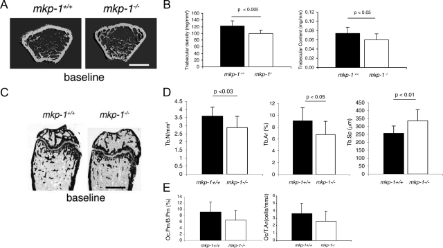
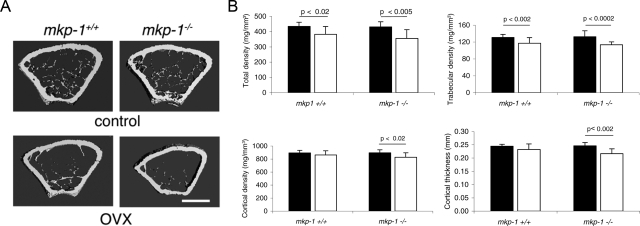
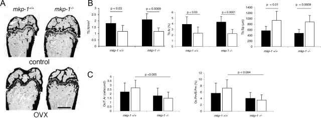
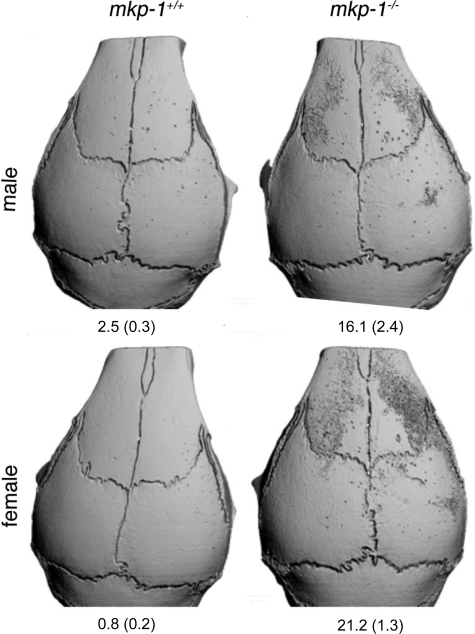
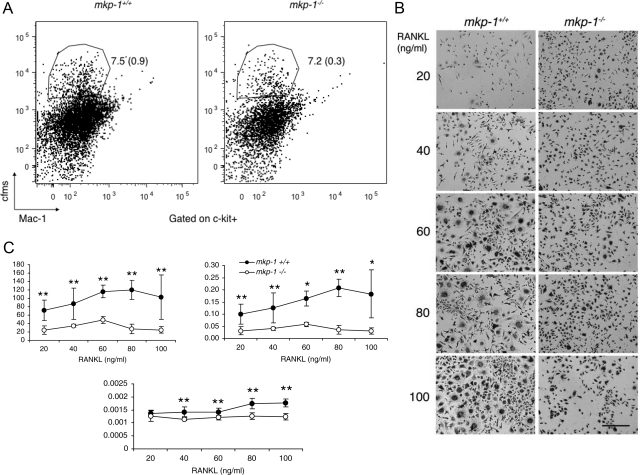
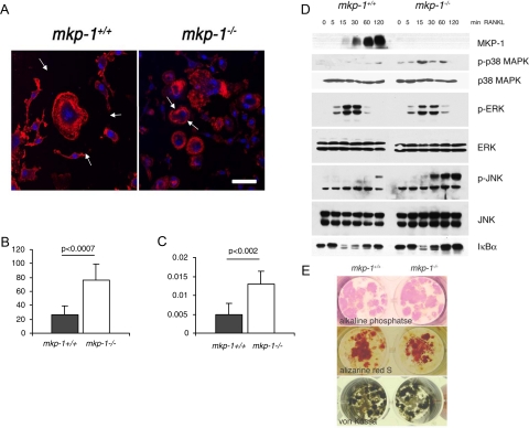
Bone mass is maintained through the complementary activities of osteoblasts and osteoclasts; yet differentiation of either osteoblasts and osteoclasts engages the mitogen-activated protein kinase (MAPK) pathway. The MAPKs are negatively regulated by a family of dual-specificity phosphatases known as the MAPK phosphatases (MKPs). MKP-1 is a stress-responsive MKP that inactivates the MAPKs and plays a central role in macrophages; however, whether MKP-1 plays a role in the maintenance of bone mass has yet to be investigated. We show here, using a genetic approach, that mkp-1(-/-) female mice exhibited slightly reduced bone mass. We found that mkp-1(+/+) and mkp-1(-/-) mice had equivalent levels of bone loss after ovariectomy despite mkp-1(-/-) mice having fewer osteoclasts, suggesting that mkp-1(-/-) osteoclasts are hyperactive. Indeed, deletion of MKP1 led to a profound activation of osteoclasts in vivo in response to local lipopolysaccharide (LPS) injection. These results suggest a role for MKP-1 in osteoclasts, which originate from the fusion of macrophages. In support of these observations, receptor activator for nuclear factor-kappaB ligand induced the expression for MKP-1, and osteoclasts derived from mkp-1(-/-) mice had increased resorptive activity. Finally, receptor activator of nuclear factor-kappaB ligand-induced p38 MAPK and c-Jun NH2-terminal kinase activities were enhanced in osteoclasts derived from mkp-1(-/-) mice. Taken together, these results show that MKP-1 plays a role in the maintenance of bone mass and does so by negatively regulating MAPK-dependent osteoclast signaling.
Figures
Similar articles
-
Diversity and specificity of the mitogen-activated protein kinase phosphatase-1 functions.Cell Mol Life Sci. 2013 Jan;70(2):223-37. doi: 10.1007/s00018-012-1041-2. Epub 2012 Jun 14. Cell Mol Life Sci. 2013. PMID: 22695679 Free PMC article. Review.
-
Caffeic acid 3,4-dihydroxy-phenethyl ester suppresses receptor activator of NF-κB ligand–induced osteoclastogenesis and prevents ovariectomy-induced bone loss through inhibition of mitogen-activated protein kinase/activator protein 1 and Ca2+–nuclear factor of activated T-cells cytoplasmic 1 signaling pathways.J Bone Miner Res. 2012 Jun;27(6):1298-1308. doi: 10.1002/jbmr.1576. J Bone Miner Res. 2012. PMID: 22337253
-
MKP-1 signaling events are required for early osteoclastogenesis in lineage defined progenitor populations by disrupting RANKL-induced NFATc1 nuclear translocation.Bone. 2014 Mar;60:16-25. doi: 10.1016/j.bone.2013.11.012. Epub 2013 Nov 20. Bone. 2014. PMID: 24269279 Free PMC article.
-
Maslinic acid suppresses osteoclastogenesis and prevents ovariectomy-induced bone loss by regulating RANKL-mediated NF-κB and MAPK signaling pathways.J Bone Miner Res. 2011 Mar;26(3):644-56. doi: 10.1002/jbmr.242. J Bone Miner Res. 2011. PMID: 20814972
-
Mitogen-activated protein kinase phosphatase-1 - a potential therapeutic target in metabolic disease.Expert Opin Ther Targets. 2010 Dec;14(12):1323-32. doi: 10.1517/14728222.2010.528395. Expert Opin Ther Targets. 2010. PMID: 21058921 Free PMC article. Review.
Cited by
-
Mitogen-activated protein kinase phosphatase-1 is a key regulator of hypoxia-induced vascular endothelial growth factor expression and vessel density in lung.Am J Pathol. 2011 Jan;178(1):98-109. doi: 10.1016/j.ajpath.2010.11.025. Epub 2010 Dec 23. Am J Pathol. 2011. PMID: 21224048 Free PMC article.
-
7-Ketocholesterol-Induced Micro-RNA-107-5p Increases Number and Activity of Osteoclasts by Targeting MKP1.Int J Mol Sci. 2022 Mar 28;23(7):3697. doi: 10.3390/ijms23073697. Int J Mol Sci. 2022. PMID: 35409056 Free PMC article.
-
Diversity and specificity of the mitogen-activated protein kinase phosphatase-1 functions.Cell Mol Life Sci. 2013 Jan;70(2):223-37. doi: 10.1007/s00018-012-1041-2. Epub 2012 Jun 14. Cell Mol Life Sci. 2013. PMID: 22695679 Free PMC article. Review.
-
Immune and inflammatory pathways are involved in inherent bone marrow ossification.Clin Orthop Relat Res. 2012 Sep;470(9):2528-40. doi: 10.1007/s11999-012-2459-4. Clin Orthop Relat Res. 2012. PMID: 22798134 Free PMC article.
-
Protein tyrosine phosphatases ε and α perform nonredundant roles in osteoclasts.Mol Biol Cell. 2014 Jun;25(11):1808-18. doi: 10.1091/mbc.E14-03-0788. Epub 2014 Apr 2. Mol Biol Cell. 2014. PMID: 24694598 Free PMC article.
References
-
- Cuevas BD, Abell AN, Johnson GL. Role of mitogen-activated protein kinase kinase kinases in signal integration. Oncogene. 2007;26:3159–3171. - PubMed
-
- Keyse SM. Dual-specificity MAP kinase phosphatases (MKPs) and cancer. Cancer Metastasis Rev. 2008;27:253–261. - PubMed
-
- Dickinson RJ, Keyse SM. Diverse physiological functions for dual-specificity MAP kinase phosphatases. J Cell Sci. 2006;119:4607–4615. - PubMed
-
- Schindeler A, Little DG. Ras-MAPK signaling in osteogenic differentiation: friend or foe? J Bone Miner Res. 2006;21:1331–1338. - PubMed
-
- Ducy P, Schinke T, Karsenty G. The osteoblast: a sophisticated fibroblast under central surveillance. Science. 2000;289:1501–1504. - PubMed
Publication types
MeSH terms
Substances
Grants and funding
LinkOut - more resources
Full Text Sources
Molecular Biology Databases
Miscellaneous
