. 2009 Nov 6;284(45):30917-24.
doi: 10.1074/jbc.M109.021238. Epub 2009 Sep 17.

Missense mutations that cause Bruck syndrome affect enzymatic activity, folding, and oligomerization of lysyl hydroxylase 2

Affiliations

Missense mutations that cause Bruck syndrome affect enzymatic activity, folding, and oligomerization of lysyl hydroxylase 2

Marjo Hyry et al. J Biol Chem. .

Abstract

Bruck syndrome is a rare autosomal recessive connective tissue disorder characterized by fragile bones, joint contractures, scoliosis, and osteoporosis. The telopeptides of bone collagen I are underhydroxylated in these patients, leading to abnormal collagen cross-linking. Three point mutations in lysyl hydroxylase (LH) 2, the enzyme responsible for the hydroxylation of collagen telopeptides, have been identified in Bruck syndrome. As none of them affects the residues known to be critical for LH activity, we studied their consequences at the molecular level by analyzing the folding and catalytic properties of the corresponding mutant recombinant polypeptides. Folding and oligomerization of the R594H and G597V mutants were abnormal, and their activity was reduced by >95% relative to the wild type. The T604I mutation did not affect the folding properties, although the mutant retained only approximately 8% activity under standard assay conditions. As the reduced activity was caused by a 10-fold increase in the K(m) for 2-oxoglutarate, the mutation interferes with binding of this cosubstrate. In the presence of a saturating 2-oxoglutarate concentration, the activity of the T604I mutant was approximately 30% of that of the wild type. However, the T604I mutant did not generate detectable amounts of hydroxylysine in the N-terminal telopeptide of a recombinant procollagen I chain when coexpressed in insect cells. The low activity of the mutant LH2 polypeptides is in accordance with the markedly reduced extent of collagen telopeptide hydroxylation in Bruck syndrome, with consequent changes in the cross-linking of collagen fibrils and severe abnormalities in the skeletal structures.


Figures

FIGURE 1.
FIGURE 1.
Schematic presentation of the locations of mutations R594H, G597V, and T604I reported in Bruck syndrome 2 relative to the catalytically critical residues in the C-terminal part of the processed LH2(long) polypeptide. His662, Asp664, and His714 correspond to the conserved residues known to be critical for binding of the Fe2+ atom in LH1 and several other 2-oxoglutarate-dependent dioxygenases, and Arg724 corresponds to the conserved basic residue required for the binding of the C-5 carboxyl group of 2-oxoglutarate. LH2(long) is a 758-amino acid polypeptide; processed LH2(long) consists of 733 amino acids after cleavage of the 25-residue signal peptide. The amino acids of LH2(long) are numbered according to the processed polypeptide.
FIGURE 2.
FIGURE 2.
Analysis of purification of the wild-type and mutant recombinant human LH2(long) polypeptides. Wild-type and mutant LH2(long) polypeptides were expressed in insect cells, and the secreted polypeptides were subjected to purification by metal chelate affinity chromatography. Aliquots of samples of the proteins bound to the column before washing (A), present in the flow-through (B) and wash (C) fractions, and remaining bound to the column after washing (D) were analyzed by 8% SDS-PAGE, followed by Coomassie Blue staining. The recombinant LH2(long) polypeptides expressed are indicated above the panels, and the positions of the wild-type and mutant LH2(long) polypeptides are indicated by the arrow.
FIGURE 3.
FIGURE 3.
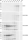
Analysis of elution of the wild-type and mutant recombinant human LH2(long) polypeptides from a metal chelate affinity column. The elution fractions were collected and analyzed by 8% SDS-PAGE, followed by Coomassie Blue staining. The elution fraction (Fr) numbers are indicated above the panels, and the positions of the wild-type and mutant LH2(long) polypeptides are indicated by arrows.
FIGURE 4.
FIGURE 4.
Analysis of the purified wild-type and mutant recombinant human LH2(long) polypeptides by SDS-PAGE under reducing conditions and by nondenaturing PAGE. The elution fractions containing the most pure wild-type and mutant LH2(long) polypeptides were pooled, concentrated, and passed through a PD-10 column to remove the histidine used in the elution, and the purified enzymes were analyzed by 6% SDS-PAGE under reducing conditions (A and B) and by 8% nondenaturing PAGE (C and D), followed by Coomassie Blue staining (A and C) or ECL Western blotting (B and D). The identity of the polypeptides is indicated above the panels, and the positions of the LH2(long) polypeptide (A and B) and the LH2(long) dimer (C and D) are indicated by arrowheads.
FIGURE 5.
FIGURE 5.
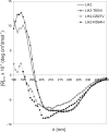
Far-UV CD spectra of the purified wild-type and mutant recombinant human LH2(long) enzymes. deg, degrees.
FIGURE 6.
FIGURE 6.
SDS-PAGE analysis of the purified wild-type and mutant recombinant human LH2(long) polypeptides after thermolysin digestion. Wild-type and mutant LH2(long) polypeptides were digested with thermolysin (TL) at a 1:500 protease/LH2 ratio and analyzed by 12% SDS-PAGE under reducing conditions, followed by Coomassie Blue staining. The identity of the polypeptides is indicated above the panel, and the position of the undigested LH2 polypeptide is indicated by the arrow.
FIGURE 7.
FIGURE 7.
Detail of the elution positions of hydroxylysine (Hyl, phenylthiohydantoin-Hyl) and lysine (K, phenylthiohydantoin-Lys) in the reverse-phase HPLC profile of the N-terminal telopeptide lysine (Lys9) of pepsin-digested type I procollagen homotrimers coexpressed with wild-type (A) or T604I mutant (B) LH2(long) in Edman degradation sequence analysis. Hydroxylysine gives two major phenylthiohydantoin peaks, one that elutes between dimethylphenylthiourea and phenylthiohydantoin-alanine and another that elutes between phenylthiohydantoin-valine and diphenylthiourea (DPTU), the latter being 10–12 times higher than the first one. The first peak was below the detection limit in our analysis. Dimethylphenylthiourea and diphenylthiourea are by-products of the sequencing reaction. mAU, milliabsorbance units.

References

    1. Kivirikko K. I., Pihlajaniemi T. (1998) Adv. Enzymol. Relat. Areas Mol. Biol. 72, 325–398 - PubMed
    1. Myllyharju J., Kivirikko K. I. (2004) Trends Genet. 20, 34–45 - PubMed
    1. Myllyharju J. (2005) Top. Curr. Chem. 247, 115–147
    1. Valtavaara M., Papponen H., Pirttilä A. M., Hiltunen K., Helander H., Myllylä R. (1997) J. Biol. Chem. 272, 6831–6834 - PubMed
    1. Passoja K., Rautavuoma K., Ala-Kokko L., Kosonen T., Kivirikko K. I. (1998) Proc. Natl. Acad. Sci. U.S.A. 95, 10482–10486 - PMC - PubMed

Publication types

MeSH terms

Substances