. 2009 Nov 4;28(21):3315-28.
doi: 10.1038/emboj.2009.267. Epub 2009 Sep 17.

Ligand-regulated oligomerization of beta(2)-adrenoceptors in a model lipid bilayer

Affiliations

Ligand-regulated oligomerization of beta(2)-adrenoceptors in a model lipid bilayer

Juan José Fung et al. EMBO J. .

Abstract

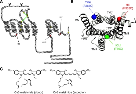
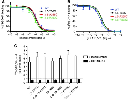
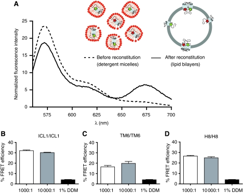
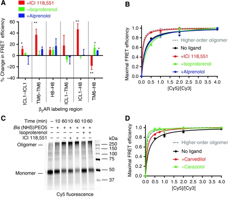
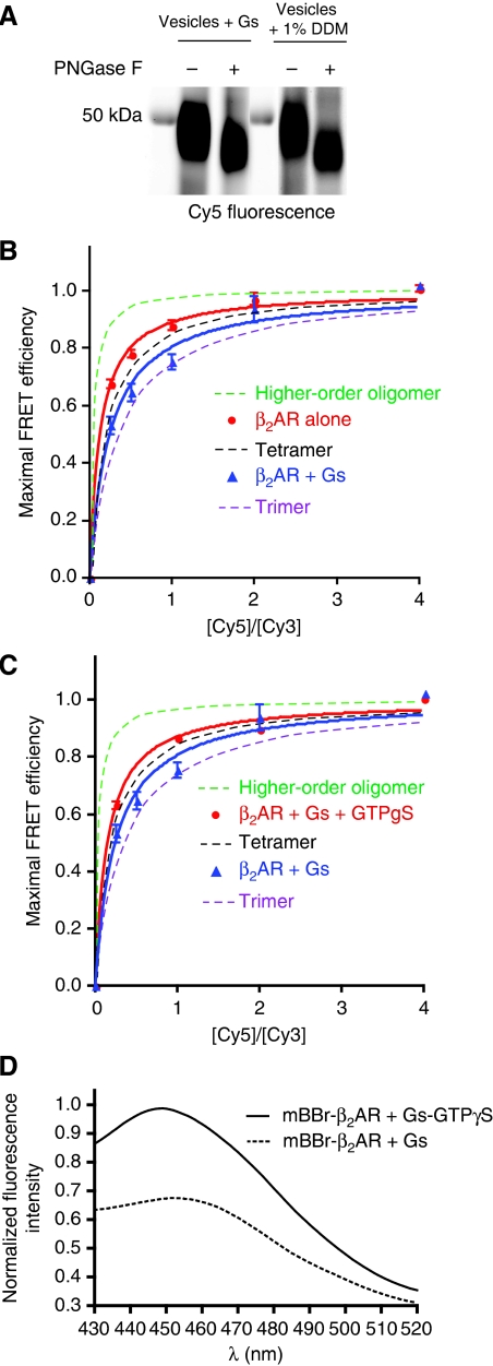
The beta(2)-adrenoceptor (beta(2)AR) was one of the first Family A G protein-coupled receptors (GPCRs) shown to form oligomers in cellular membranes, yet we still know little about the number and arrangement of protomers in oligomers, the influence of ligands on the organization or stability of oligomers, or the requirement for other proteins to promote oligomerization. We used fluorescence resonance energy transfer (FRET) to characterize the oligomerization of purified beta(2)AR site-specifically labelled at three different positions with fluorophores and reconstituted into a model lipid bilayer. Our results suggest that the beta(2)AR is predominantly tetrameric following reconstitution into phospholipid vesicles. Agonists and antagonists have little effect on the relative orientation of protomers in oligomeric complexes. In contrast, binding of inverse agonists leads to significant increases in FRET efficiencies for most labelling pairs, suggesting that this class of ligand promotes tighter packing of protomers and/or the formation of more complex oligomers by reducing conformational fluctuations in individual protomers. The results provide new structural insights into beta(2)AR oligomerization and suggest a possible mechanism for the functional effects of inverse agonists.


Conflict of interest statement

The authors declare that they have no conflict of interest.

Figures

Figure 1
Figure 1
β2AR single-cysteine constructs and FRET donor–acceptor pair. (A) Three single-reactive cysteines constructs were generated on a minimal cysteine background (Δ5-β2AR). The labelling sites were placed in the first ICL, Δ5-β2AR-T66C, at the cytoplasmic end of the sixth transmembrane segment, Δ5-β2AR-A265C, and helix eight, Δ5-β2AR-R333C. (B) Intracellular 3D view of the distribution of regions chosen for single-cysteine mutants, α-carbons are depicted. (C) FRET donor (λex=549 nm; λem=570 nm) and acceptor pair (λex=650 nm; λem=670 nm).
Figure 2
Figure 2
β2ARs are predominantly oriented outside-out in lipid bilayers. (A) Strategies for determining orientation of β2AR in lipid bilayers. (B) Purified receptors were reconstituted as described under Materials and methods and then subjected to treatment with Factor Xa and resolved by 10% SDS–PAGE and transferred onto nitrocellulose. The presence of β2AR was determined by probing with an M1 antibody conjugated with Alexa-680. (C) Samples subjected to PNGase F were prepared and imaged as in panel A. (D, E) Reconstituted samples were treated with the hydrophilic, amine-reactive, alkylating reagent NHS-PEO4-biotin that disrupts binding of the M1 monoclonal antibody to the FLAG epitope. Samples were assessed for reactivity to M1 antibody (D) and an antibody that recognizes the C-terminal six-histidine tag (E). All data are representative of three independent experiments.
Figure 3
Figure 3
β2ARs are homogenously distributed in lipid vesicles. (A) To determine the distribution of β2ARs in lipid vesicles, sucrose density gradients of samples containing 0.4% NBD–phosphocholine and Cy5–β2ARs reconstituted at a lipid-to-receptor ratio of 1000:1 were performed as described in the Supplementary data. Detection of lipid fractions was performed by following NBD fluorescence (λex=460 nm) and receptor fractions by following Cy5 fluorescence (λex=649 nm). (B) Reconstituted β2ARs were imaged using a negative staining protocol as described in the Supplementary data to determine the size distribution of vesicles and the number of receptors per vesicle. Scale bar length represents 200 nm. Data are representative of three independent experiments.
Figure 4
Figure 4
Single-reactive cysteine mutants are fully functional. The affinity of the agonist isoproterenol (A) and the inverse agonist ICI 118,551 (B) was measured for all three single-cysteine mutants (Δ5-T66C, Δ5-A265C and Δ5-R333C) and wild-type receptor by competitive binding of [3H]-DHA. Results are expressed as percent of radio-ligand bound in the absence of competitor. (C) Functionality of the three single-cysteine mutants, unlabelled or labelled with Cy5, and wild-type receptor was determined by GTPγS binding as described in the Supplementary data. [35S]-GTPγS-specific binding induced by 10 μM isoproterenol (agonist response) or by 10 μM ICI 118,551 (inverse agonist response) is shown as fold over basal. All functional data represent the mean±s.e.m. of three independent experiments performed in triplicate.
Figure 5
Figure 5
Intermolecular FRET between Cy3- and Cy5-labelled β2AR is independent of other cellular proteins and is specific. (A) Purified, detergent-solubilized receptor protein was labelled with Cy3 or Cy5 maleimide and unreacted fluorophore was quenched with cysteine and separated from protein by gel filtration as described under Materials and methods. Cy3- and Cy5-labelled protein samples were mixed at a 1:1 molar ratio and reconstituted into phospholipids bilayers or maintained in detergent. Subtraction of the proper controls and normalization of the raw traces is described in the Supplementary data. Labelled β2ARs were reconstituted at a 10-fold higher lipid-to-receptor ratio (10 000:1) and FRET efficiency was measured for ICL1/ICL1 (B), TM6/TM6 (C) and H8/H8 (D) interactions. Data are representative of at least three independent experiments (A) or represent the mean±s.e.m. of at least three independent experiments (B–D).
Figure 6
Figure 6
Specificity of β2AR oligomerization as assessed by FRET saturation. FRET saturation involved varying the ratio of Cy5- to Cy3-labelled β2ARs over a range of 1:1 to 10:1 (Cy5:Cy3), while the overall β2AR concentration was kept constant. Saturable FRET is observed for ICL1/ICL1 (A), TM6/TM6 (B) and H8/H8 (C). FRET measurements were performed and calculated as described in the Supplementary data. Data represent the mean±s.e.m. of at least three independent experiments. (D) FRET saturation data from all three constructs (A–C above) was normalized to maximal FRET efficiency and then averaged and plotted together with theoretical curves (dashed lines) for dimer, trimer, tetramer and higher-order oligomer that were generated using equation (1) in the Supplementary data.
Figure 7
Figure 7
β2AR oligomers are regulated by inverse agonists. (A) Treatment of FRET samples with saturating amounts of the inverse agonist ICI 118,551, agonist isoproterenol and neutral antagonist alprenolol. (B) FRET saturation in the presence of ligands. Isoproterenol and alprenolol led to no observable difference from the unliganded FRET saturation curve, whereas ICI 118,551 yielded to a curve that is more consistent with higher-order oligomers. (C) Cross-linking of reconstituted Cy5-labelled β2AR samples in the presence or absence of isoproterenol or ICI 118,551 was carried out as described in the Supplementary data. (D) FRET saturation in the presence of the inverse agonists carvedilol (red) and carazolol (green). All data are reported as mean±s.e.m. (A, B, D) or are representative of at least three independent experiments (C). * (P<0.05) and ** (P<0.005).
Figure 8
Figure 8
Effect of the G protein Gs on FRET saturation of Cy5- and Cy3-labelled β2AR. FRET saturation was performed by varying the ratio of Cy5- to Cy3-labelled β2AR-R333C over a range of 1:4 to 4:1 (Cy5:Cy3), while the overall β2AR concentration was kept constant. Purified Gs heterotrimer was added at a molar ratio of 3 Gs:1 β2AR before reconstitution. (A) The inclusion of Gs in the reconstitution did not alter the orientation of β2AR in vesicles as determined by the susceptibility of reconstituted β2AR to PNGase F (see Figure 3C). FRET saturation was significantly lower in the presence of Gs compared with β2AR alone (B) or β2AR and Gs with 10 μM GTPγS (C). (D) β2AR was labelled on C265 at the cytoplasmic end of TM6 with mBBr–β2AR and reconstituted with Gs under the same conditions that were used for FRET saturation experiments. Gs induced a decrease in intensity and a 4-nm shift in λMAX of mBBr–β2AR relative to the same reconstitution in the presence of GTPγS. A two-way ANOVA was used to compare FRET values for β2AR, β2AR+Gs and β2AR+Gs+GTPγS at the different Cy5:Cy3 ratios. A posteriori statistical analysis showed significant decrease in FRET between β2AR and β2AR+Gs (P<0.008), and a significant increase in FRET between β2AR+Gs and β2AR+Gs+GTPγS (P<0.04) for all Cy5:Cy3 ratios except 2 and 4. No statistical differences are found between β2AR and β2AR+Gs+GTPγS.
Figure 9
Figure 9
Schematic representation of possible β2AR oligomers. (A) Cytoplasmic view of the 3D structure of the β2AR (left) and cartoon of this footprint (right), with the centre of mass of Cy3 depicted as spheres: T66C (green), A265C (blue) and R333C (red). TM6 is depicted in the inactive conformation (top) and in the proposed active conformation as observed in the structure of opsin (bottom). (B) Receptor oligomerization involving the surfaces of TMs 4 or/and 5 is not compatible with our FRET results and might sterically prevent movement of TM6. (C) Our FRET results suggest an arrangement of protomers involving the TM1 interface. (D) The movement of the cytoplasmic end of TM6 upon agonist binding repositions the fluorophore outwards in two of the four protomers, and towards H8 in the other two. (E) Treatment with the inverse agonist ICI probably reduces conformational fluctuations responsible for basal activity, increasing the packing of the oligomers. (F) ICI may stabilize higher-order oligomers where TM6 is packed into the core of the oligomer, contributing to increase FRET efficiency and possibly the inactive state of the receptor.

References

    1. Allen JA, Halverson-Tamboli RA, Rasenick MM (2007) Lipid raft microdomains and neurotransmitter signalling. Nat Rev Neurosci 8: 128–140 - PubMed
    1. Altenbach C, Kusnetzow AK, Ernst OP, Hofmann KP, Hubbell WL (2008) High-resolution distance mapping in rhodopsin reveals the pattern of helix movement due to activation. Proc Natl Acad Sci USA 105: 7439–7444 - PMC - PubMed
    1. Angers S, Bouvier M (2000) Reply: beyond receptor dimerization. Trends Pharmacol Sci 21: 326. - PubMed
    1. Angers S, Salahpour A, Joly E, Hilairet S, Chelsky D, Dennis M, Bouvier M (2000) Detection of beta 2-adrenergic receptor dimerization in living cells using bioluminescence resonance energy transfer (BRET). Proc Natl Acad Sci USA 97: 3684–3689 - PMC - PubMed
    1. Bayburt TH, Leitz AJ, Xie G, Oprian DD, Sligar SG (2007) Transducin activation by nanoscale lipid bilayers containing one and two rhodopsins. J Biol Chem 282: 14875–14881 - PubMed

Publication types