

Case Reports
doi: 10.3340/jkns.2009.46.2.152.
Epub 2009 Aug 31.
Intradural retroclival chordoma
Affiliations
- PMID: 19763218
- PMCID: PMC2744025
- DOI: 10.3340/jkns.2009.46.2.152
Item in Clipboard
Case Reports
Intradural retroclival chordoma
J Korean Neurosurg Soc.
2009 Aug.
Abstract
A 43-year-old woman presented with dizziness, ataxia and right hearing difficulty. Her magnetic resonance images demonstrated an inhomogeneously contrast-enhanced large tumor growing into right cavernous sinus and Meckel's cave located totally within intradural retroclival region. She underwent retromastoid suboccipital craniotomy to resect the tumor mass and adjuvant gamma knife radiosurgery for remnant tumor at 1 month after operation. Adjuvant radiosurgery after surgical excision seems to be effective for the treatment of intradural extraosseous chordomas.
Keywords: Chordoma; Intradural; Retroclival.
Figures
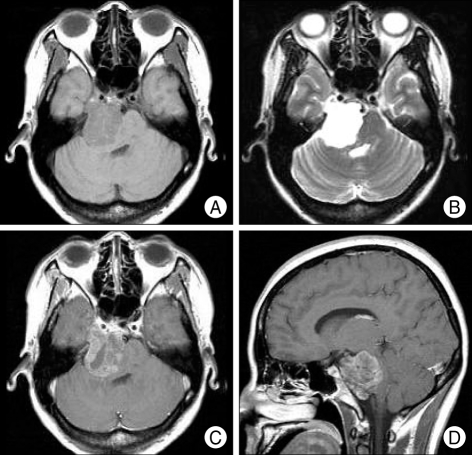
Preoperative MR images presenting the intradural retroclival tumor with growing into right cavernous sinus and Meckel's cave. A : Axial T1-weigted imgae. B : Axial T-2 weighted image. C : Axial Gd-enhanced T1-weighted image. D : Sagittal Gd-enhanced T1-weighted image.
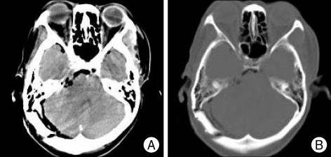
Postoperative brain CT demonstrates no definite contrast enhancing mass in right cerebellopontine angle region and no bony destruction. A : Axial contrast enhanced brain CT image. B : Bone-setting image.
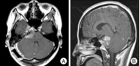
Follow-up MR images at 1 month after operation demonstrates a remnant tumor. A : Axial Gd-enhanced T1-weighted image. B : Sagittal Gd-enhanced T1-weighted image.
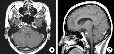
MR images, performed 14 months after gammaknife radiosurgery, representing decreased remnant tumor in size. A : Axial Gd-enhanced T1-weighted image. B : Sagittal Gd-enhanced T1-weighted image.
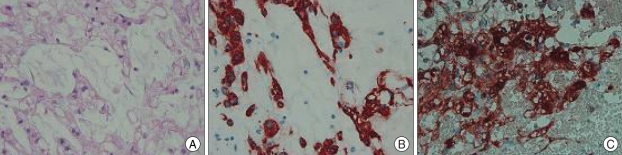
Photomicrograph of the intradural tumor showing typical physaliphorous cells in mucinous matrix. H & E stain ×200 (A). Immunohistochemical stains showing expression of cytokeratin (B), and S-100 protein (C).
References
-
- Bartolini G. [Cordoma del clivus quale causa di emorragia cerebrale : discussione anatomo-clinica su di una osservazione autoptica] Boll Soc Ital Biol Sper. 1974;50:912–918. - PubMed
-
- Dahlin DC, MacCarty CS. Chordoma. Cancer. 1952;5:1170–1178. - PubMed
-
- Dow GR, Robson DK, Jaspan T, Punt JA. Intradural cerebellar chordoma in a child : a case report and review of the literature. Childs Nerv Syst. 2003;19:188–191. - PubMed
-
- Inci S, Palaoglu S, Önol B, Erbengi A. Low cervical chordoma: case report. Spinal Cord. 1996;34:358–360. - PubMed
Publication types
LinkOut - more resources
Full Text Sources
