. 2009 Sep 18;4(9):e7076.
doi: 10.1371/journal.pone.0007076.

Transcriptional signature and memory retention of human-induced pluripotent stem cells

Affiliations

Transcriptional signature and memory retention of human-induced pluripotent stem cells

Maria C N Marchetto et al. PLoS One. .

Abstract

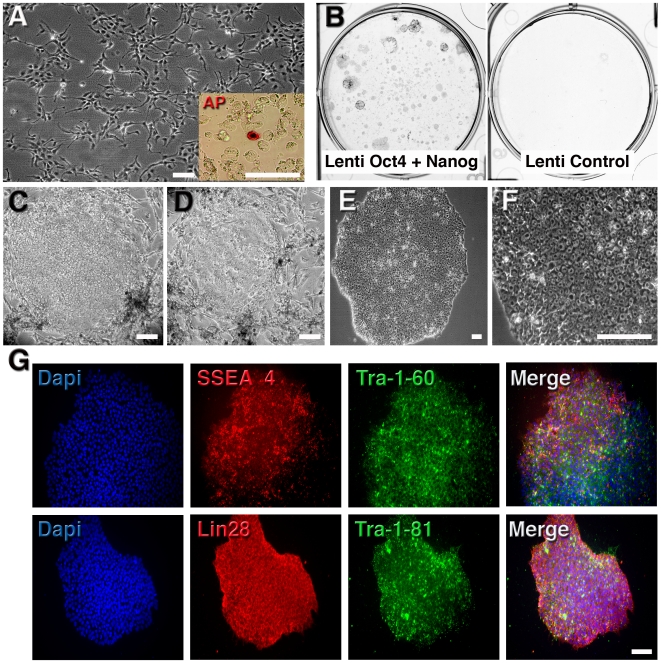
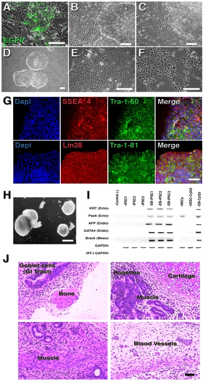
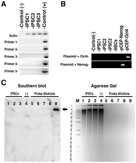
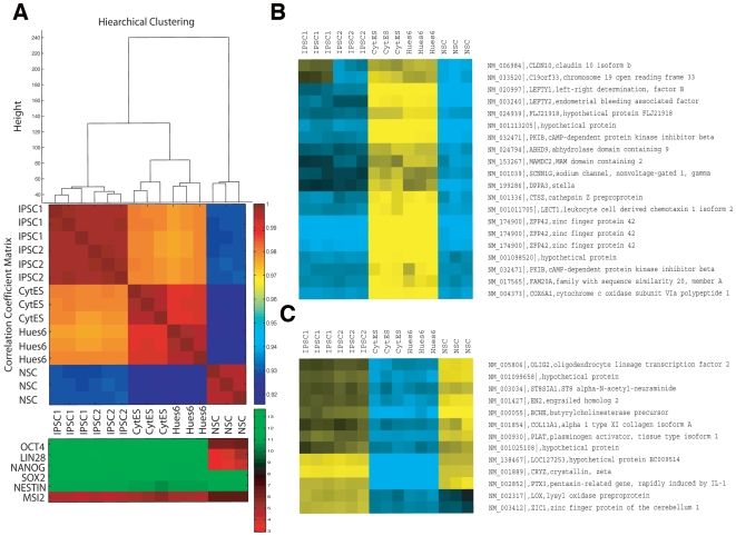
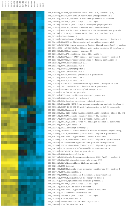
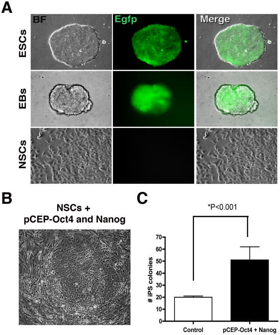
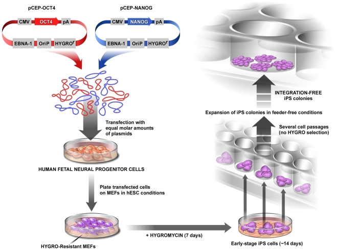
Genetic reprogramming of somatic cells to a pluripotent state (induced pluripotent stem cells or iPSCs) by over-expression of specific genes has been accomplished using mouse and human cells. However, it is still unclear how similar human iPSCs are to human Embryonic Stem Cells (hESCs). Here, we describe the transcriptional profile of human iPSCs generated without viral vectors or genomic insertions, revealing that these cells are in general similar to hESCs but with significant differences. For the generation of human iPSCs without viral vectors or genomic insertions, pluripotent factors Oct4 and Nanog were cloned in episomal vectors and transfected into human fetal neural progenitor cells. The transient expression of these two factors, or from Oct4 alone, resulted in efficient generation of human iPSCs. The reprogramming strategy described here revealed a potential transcriptional signature for human iPSCs yet retaining the gene expression of donor cells in human reprogrammed cells free of viral and transgene interference. Moreover, the episomal reprogramming strategy represents a safe way to generate human iPSCs for clinical purposes and basic research.


Conflict of interest statement

Competing Interests: The authors have declared that no competing interests exist.

Figures

Figure 1
Figure 1. Efficient and rapid generation of iPSCs from human fetal NSCs using two factors.
A, Morphology of human fetal NSCs before lentiviral infection. Inset: after 3 days post-infection with Lenti-Oct4 and Lenti-Nanog, individual cells expressed alkaline phosphatase (AP). B, Example of infected plates stained for AP at 14 days post-infection showing several AP-positive colonies. Control infection did not result in any AP-positive colonies. C and D, Aspect of colonies 14 days after infection growing in MEFs. E, Established human iPSC colonies, with well-defined borders and compact cells, are morphologically similar to hESCs. F, Typical image of iPSCs growing in feeder-free conditions. G, Representative immunofluorescence analysis of iPSCs growing on matrigel. Clear expression of pluripotent markers is observed. Bar = 150 µm.
Figure 2
Figure 2. Generation of virus-free, integration-free human iPSCs.
A, Aspect of human NSCs after plasmid electroporation and plating on MEFs. B and C, Some selected colonies display a strong differentiation tendency in feeder-free conditions. D, Established iPSC lines are morphologically similar to hESCs. E, iPSCs have a large nucleus-to-cytoplasm ratio and prominent nucleoli when compared to original NSCs (F). G, Immunofluorescence analysis of iPSCs growing on matrigel showed clear expression of typical ESC markers. H, In vitro differentiation of iPSCs into EBs. I, RT-PCR from undifferentiated and EB-derived iPSCs showing expression of markers for all three primary germ cell layers. The hESCs Cyt25 was used as a benchmark. J, Hematoxylin and eosin staining of teratoma sections generated from integration-free iPSC lines showing differentiation in three germ layers: goblet cells in gastro-intestinal (GI) tract (endoderm); neural rosettes (ectoderm) and blood vessels, muscle and cartilage/bone (mesoderm). Bar = 150 µm.
Figure 3
Figure 3. Absence of plasmid integration on virus-free iPSCs.
A and B, PCR analyses for plasmid integration in genomic DNA from the iPSC clones. Controls: (−) water; (+) pCEP4 plasmid. Primers were designed to specifically amplify plasmid backbone (A) or transgenes (B) (see Methods). c, Southern blot (left) membrane hybridization of 10 µg of BamHI-digested genomic DNA (see corresponding agarose gel on right) using a DNA probe from the pCEP backbone. Plasmid DNAs of pCEP-Oct4 and PCEP-Nanog, diluted to the equivalent of 0.5 integration per genome, were used as controls for probe dilution. Lanes: M, DNA molecular marker; 1- iPSC1; 2- iPSC2; 3- iPSC3; 4- NSCs (negative control); 5- probe 25 ρg; 6- probe 50 ρg; 7- 100 ρg; 8- 200 ρg and 9- 50 ηg. Arrow indicates expected probe size.
Figure 4
Figure 4. Transcriptional analysis of human integration-free iPSC colonies.
A, Hierarchical clustering and correlation coefficients of microarray profiles of triplicate iPSC1, iPSC2, CytES (Cyt25 hESC), Hues6 and NSC. Color bar indicates the level of correlation (from 0 to 1). Panel below illustrates marker genes implicated in pluripotency of NSCs, with color bar reporting log2 normalized expression values (green/red indicates high/low relative expression). B, Refseq-annotated genes that were insufficiently induced in iPSCs relative to hESCs (yellow/blue indicates high normalized log2 expression). C, Refseq-annotated genes that were insufficiently silenced in iPSCs relative to hESCs.
Figure 5
Figure 5. Refseq-annotated genes that were upregulated in iPSCs relative to both hESCs and NSC.
Panel illustrates marker genes implicated in pluripotency of NSCs, with color bar reporting log2 normalized expression values (green/red indicates high/low relative expression).
Figure 6
Figure 6. The dynamics of integration-free reprogramming.
A, Undifferentiated H1 Oct4-EGFP hESC line expresses the EGFP reporter gene that is gradually turned off during NSC differentiation. NSCs are morphologically distinct from hESCs. B, Small iPSC colonies can be detected 10 days after transfection with pCEP-Oct4 and pCEP-Nanog. C, Typical number of iPSC colonies obtained with electroporation of pCEP-Oct4 and Nanog or with Oct4 alone. Bar = 150 µm.
Figure 7
Figure 7. Schematic model of integration-free human iPSC generation from NSCs.
Episomal plasmids carrying reprogramming factors are transfected into NSCs and cells are plated on MEFs. On the following day, medium is changed to the hESC condition. Resistant selection is kept for a week. After 14 days, iPSC colonies are visible and can be transferred to a feeder-free condition. Individual colonies are expanded and ready for characterization. At this time, no evidence of plasmid integration is found.

References

    1. Takahashi K, Yamanaka S. Induction of pluripotent stem cells from mouse embryonic and adult fibroblast cultures by defined factors. Cell. 2006;126:663–676. - PubMed
    1. Takahashi K, Tanabe K, Ohnuki M, Narita M, Ichisaka T, et al. Induction of pluripotent stem cells from adult human fibroblasts by defined factors. Cell. 2007;131:861–872. - PubMed
    1. Yu J, Vodyanik MA, Smuga-Otto K, Antosiewicz-Bourget J, Frane JL, et al. Induced pluripotent stem cell lines derived from human somatic cells. Science. 2007;318:1917–1920. - PubMed
    1. Lowry WE, Richter L, Yachechko R, Pyle AD, Tchieu J, et al. Generation of human induced pluripotent stem cells from dermal fibroblasts. Proc Natl Acad Sci U S A. 2008;105:2883–2888. - PMC - PubMed
    1. Park IH, Lerou PH, Zhao R, Huo H, Daley GQ. Generation of human-induced pluripotent stem cells. Nat Protoc. 2008;3:1180–1186. - PubMed

Publication types