Review
. 2009 Nov;66(22):3583-94.
doi: 10.1007/s00018-009-0145-9. Epub 2009 Sep 10.

Emerging concepts in acute mountain sickness and high-altitude cerebral edema: from the molecular to the morphological

Affiliations
Review

Emerging concepts in acute mountain sickness and high-altitude cerebral edema: from the molecular to the morphological

Damian Miles Bailey et al. Cell Mol Life Sci. 2009 Nov.

Abstract

Acute mountain sickness (AMS) is a neurological disorder that typically affects mountaineers who ascend to high altitude. The symptoms have traditionally been ascribed to intracranial hypertension caused by extracellular vasogenic edematous brain swelling subsequent to mechanical disruption of the blood-brain barrier in hypoxia. However, recent diffusion-weighted magnetic resonance imaging studies have identified mild astrocytic swelling caused by a net redistribution of fluid from the "hypoxia-primed" extracellular space to the intracellular space without any evidence for further barrier disruption or additional increment in brain edema, swelling or pressure. These findings and the observation of minor vasogenic edema present in individuals with and without AMS suggest that the symptoms are not explained by cerebral edema. This has led to a re-evaluation of the relevant pathogenic events with a specific focus on free radicals and their interaction with the trigeminovascular system.


Figures
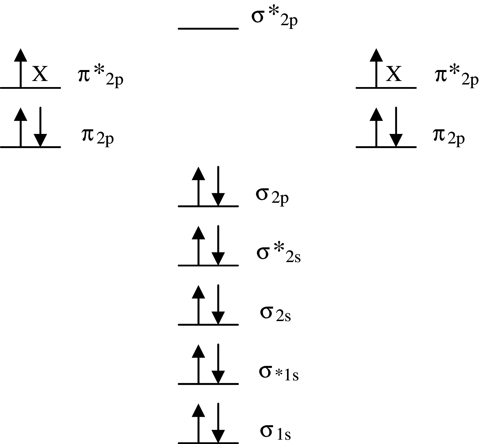
Fig. 1
Fig. 1
Molecular orbital diagram of the electronic ground state (most stable form) diatomic oxygen molecule (3∑gO2). Each line represents a molecular orbital and the arrows represent electrons, the direction of which indicates their spin quantum number. Note that O2 qualifies as a di-radical since it contains two unpaired electrons each occupying different π*2p anti-bonding orbitals (each missing electron is marked X). These unpaired electrons have the same spin quantum number (parallel spin). During the process of oxidation, O2 thermodynamically prefers to accept only one electron at a time into each of the vacant π*2p anti-bonding orbitals to conform with the Pauli Exclusion Principle. Fortuitously, this “spin restriction” means that O2 does not react vigorously with the brain’s organic compounds and instead prefers to react with other radicals
Fig. 2
Fig. 2
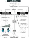
The healthy human brain pivots delicately between a bi-modal distribution of free radicals poised to increase in both hypoxia (that serve to defend cerebral oxygenation) and hyperoxia (capable of causing structural damage) subsequent to mitochondrial superoxide (O2−•) formation. The rate of increase in O2−• increases in hypoxia (primarily the Q cycle) and hyperoxia due to an increase in the concentration of electron donors (R) and O2 concentration, respectively. The resting intracellular partial pressure of oxygen is based on measurements recently documented in resting normoxic human skeletal muscle [70]
Fig. 3
Fig. 3
Normal response of the cerebral circulation to hypoxia. T 2 rt T2 relaxation time, ADC apparent diffusion co-efficient
Fig. 4
Fig. 4
3T-T2 and diffusion-weighted (DW) images of a subject with clinical AMS. Note the lack of any visual difference between normoxia and following 6 h passive exposure to hypoxia (12%O2) indicating that the edema detected by MRI is very subtle indeed. Baumgartner et al. (unpublished findings) based on [36]
Fig. 5
Fig. 5
Recent evidence suggesting that subjects diagnozed with clinical acute mountain sickness (AMS+) do not exhibit any additional increment in brain swelling (a), (vasogenic) edema (b), alteration in blood–brain barrier (BBB) permeability (c), or lumbar pressure (d). Data modified from [34]; values represent mean ± SD calculated as the difference between hypoxia relative to the normoxic control
Fig. 6
Fig. 6
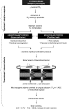
Revised schema of events implicated in the pathophysiology of acute mountain sickness (AMS) and high-altitude cerebral edema (HACE). The traditional model (A) (stippled lines) contends that vasogenic edematous brain swelling combined with a reduced CSF buffering capacity may predispose to intracranial hypertension and thus by consequence, AMS. The revised model (B) suggests that AMS is not associated with any “additional” volume overload thus arguing against a role for intracranial hypertension. It describes how free radicals can directly activate the trigeminovascular system to trigger neurovascular headache and AMS. HACE may reflect the more extreme spectrum of “osmotic-oxidative stress” resulting in gross barrier dysfunction and cerebral capillary “stress failure”. ICV Intracranial volume, ECS/ICS extra/intra-cellular space
Fig. 7
Fig. 7
X-band electron paramagnetic (EPR) spectroscopic detection of a hydroxyl (OH: a N = a H = 14.9 Gauss) and b mixture of lipid-derived alkyl (LC: a 1N = 15.8G, a 1H = 2.8G), OCH3 (a 2N = 14.5G, a 2H = 2.1G) and nitroxide (a 3N = 16.0G) free radicals in the cerebrospinal fluid of one subject in normoxia without acute mountain sickness (AMS−) and following 18 h exposure to hypoxia (12%O2) when severe acute mountain sickness (AMS+) was diagnozed. EPR spectroscopy is considered the most sensitive, specific, and direct molecular technique for the detection and subsequent identification of free radicals sine qua non [17]. The spectra illustrated were obtained using an ex vivo spin-trapping technique [34]. The spin-traps 5,5-dimethylpyrroline-N-oxide (DMPO-1 mM) and α-(4-pyridyl 1-oxide)-N-tert-butylnitrone (POBN-50 mM) were employed to detect proximal OH and distal lipid-derived species, respectively. Note the marked increase in free radical-mediated lipid peroxidation in AMS+
Fig. 8
Fig. 8
Susceptibility-weighted T2*-images (a) obtained in one subject 7 weeks after CT diagnosis of HACE following ascent to 3,860 m and shortly following clinical diagnosis of severe AMS in a separate subject at 5,895 m. Note the multiple patchy hypo-intensities (arrows) present in the HACE subject only that correspond to micro-hemorrhages that result in hemosiderin [insoluble Fe(III) oxide hydroxide] deposition secondary to blood–brain barrier disruption and cerebral capillary “stress failure”. These “micro-bleeds” were located predominantly in the genu and splenium of the corpus callosum (b) which have previously been identified as predilection sites for vasogenic edema in HACE [2]. Data modified from [69]

Similar articles

Cited by

References

    1. Hackett PH, Roach RC. High-altitude illness. N Engl J Med. 2001;345:107–114. doi: 10.1056/NEJM200107123450206. - DOI - PubMed
    1. Hackett PH, Yarnell PR, Hill R, Reynard K, Heit J, McCormick J. High-altitude cerebral edema evaluated with magnetic resonance imaging: clinical correlation and pathophysiology. J Am Med Assoc. 1998;280:1920–1925. doi: 10.1001/jama.280.22.1920. - DOI - PubMed
    1. Bartsch P, Bailey DM, Berger MM, Knauth M, Baumgartner RW. Acute mountain sickness; controversies, advances and future directions. High Alt Med Biol. 2004;5:110–124. doi: 10.1089/1527029041352108. - DOI - PubMed
    1. Roach RC, Hackett PH. Frontiers of hypoxia research: acute mountain sickness. J Exp Biol. 2001;204:3161–3170. - PubMed
    1. Basnyat B, Murdoch DR. High altitude illness. Lancet. 2003;361:1967–1974. doi: 10.1016/S0140-6736(03)13591-X. - DOI - PubMed

MeSH terms

LinkOut - more resources