

Anionic phospholipids differentially regulate the epithelial sodium channel (ENaC) by interacting with alpha, beta, and gamma ENaC subunits
- PMID: 19763606
- PMCID: PMC2871249
- DOI: 10.1007/s00424-009-0733-4
Anionic phospholipids differentially regulate the epithelial sodium channel (ENaC) by interacting with alpha, beta, and gamma ENaC subunits
Abstract
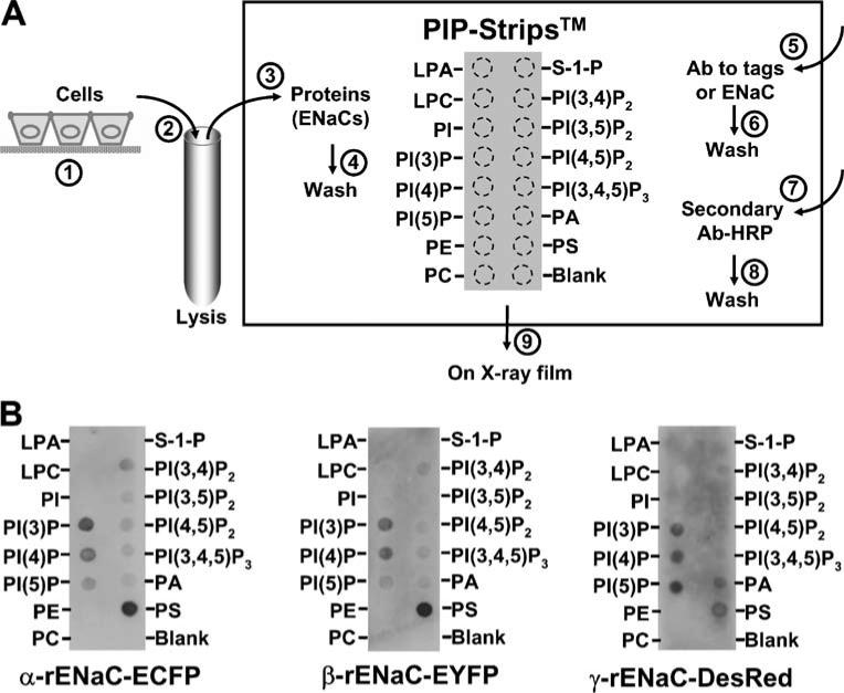
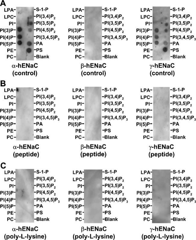
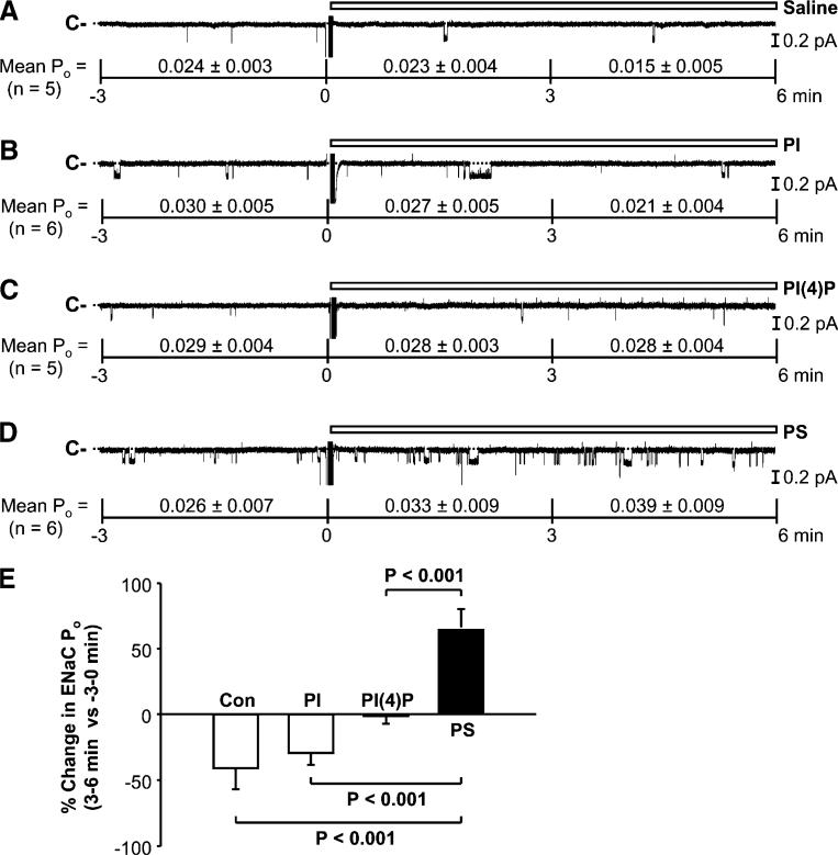
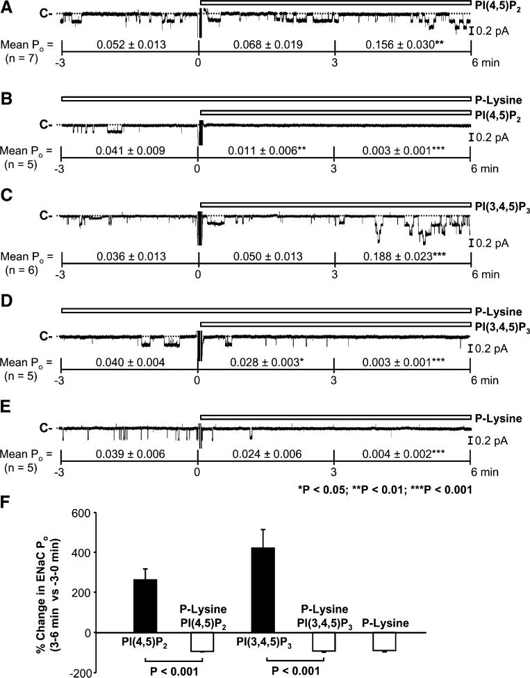
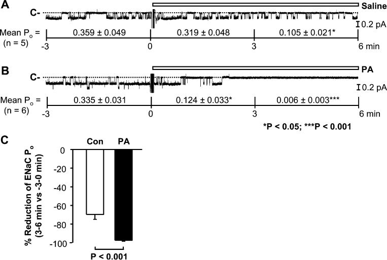
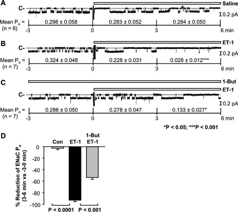
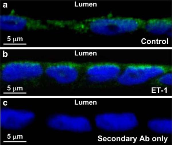
Anionic phospholipids (APs) present a variety of lipids in the cytoplasmic leaflet of the plasma membrane, including phosphatidylinositol (PI), PI-4-phosphate (PI(4)P), phosphatidylserine (PS), PI-4,5-bisphosphate (PI(4,5)P(2)), PI-3,4,5-trisphosphate (PI(3,4,5)P(3)), and phosphatidic acid (PA). We previously showed that PI(4,5)P(2) and PI(3,4,5)P(3) upregulate the renal epithelial sodium channel (ENaC). Further studies from others suggested that PI(4,5)P(2) and PI(3,4,5)P(3) respectively target beta- and gamma-ENaC subunit. To determine whether PI(4,5)P(2) and PI(3,4,5)P(3) selectively bind to beta and gamma subunit, we performed lipid-protein overlay experiments. Surprisingly, the results reveal that most APs, including PI(4)P, PS, PI(4,5)P(2), PI(3,4,5)P(3), and PA, but not PI, non-selectively bind to not only beta and gamma but also alpha subunit. To determine how these APs regulate ENaC, we performed inside-out patch-clamp experiments and found that PS, but not PI or PI(4)P, maintained ENaC activity, that PI(4,5)P(2) and PI(3,4,5)P(3) stimulated ENaC, and that PA, however, inhibited ENaC. These data together suggest that APs differentially regulate ENaC by physically interacting with alpha-, beta-, and gamma-ENaC. Further, the data from cell-attached patch-clamp and confocal microscopy experiments indicate that PA, a product of phospholipase D, may provide one of the pathways for inhibition of ENaC by endothelin receptors.
Figures
Similar articles
-
Anionic phospholipids regulate native and expressed epithelial sodium channel (ENaC).J Biol Chem. 2002 Mar 8;277(10):7641-4. doi: 10.1074/jbc.C100737200. Epub 2002 Jan 23. J Biol Chem. 2002. PMID: 11809744
-
Molecular determinants of PI(4,5)P2 and PI(3,4,5)P3 regulation of the epithelial Na+ channel.J Gen Physiol. 2007 Oct;130(4):399-413. doi: 10.1085/jgp.200709800. J Gen Physiol. 2007. PMID: 17893193 Free PMC article.
-
Acute regulation of epithelial sodium channel by anionic phospholipids.J Am Soc Nephrol. 2005 Nov;16(11):3182-7. doi: 10.1681/ASN.2005040434. Epub 2005 Sep 28. J Am Soc Nephrol. 2005. PMID: 16192420 Review.
-
Regulation of the epithelial sodium channel by phosphatidylinositides: experiments, implications, and speculations.Pflugers Arch. 2007 Oct;455(1):169-80. doi: 10.1007/s00424-007-0294-3. Epub 2007 Jun 29. Pflugers Arch. 2007. PMID: 17605040 Review.
-
Steroids and exogenous gamma-ENaC subunit modulate cation channels formed by alpha-ENaC in human B lymphocytes.J Biol Chem. 2004 Aug 6;279(32):33206-12. doi: 10.1074/jbc.M405455200. Epub 2004 Jun 8. J Biol Chem. 2004. PMID: 15187080
Cited by
-
Hydrogen peroxide stimulates the epithelial sodium channel through a phosphatidylinositide 3-kinase-dependent pathway.J Biol Chem. 2011 Sep 16;286(37):32444-53. doi: 10.1074/jbc.M111.254102. Epub 2011 Jul 27. J Biol Chem. 2011. PMID: 21795700 Free PMC article.
-
Hydrogen sulfide prevents hydrogen peroxide-induced activation of epithelial sodium channel through a PTEN/PI(3,4,5)P3 dependent pathway.PLoS One. 2013 May 31;8(5):e64304. doi: 10.1371/journal.pone.0064304. Print 2013. PLoS One. 2013. PMID: 23741314 Free PMC article.
-
Palmitate Stimulates the Epithelial Sodium Channel by Elevating Intracellular Calcium, Reactive Oxygen Species, and Phosphoinositide 3-Kinase Activity.Oxid Med Cell Longev. 2018 Dec 2;2018:7560610. doi: 10.1155/2018/7560610. eCollection 2018. Oxid Med Cell Longev. 2018. PMID: 30622672 Free PMC article.
-
ENaC activity and expression is decreased in the lungs of protein kinase C-α knockout mice.Am J Physiol Lung Cell Mol Physiol. 2014 Sep 1;307(5):L374-85. doi: 10.1152/ajplung.00040.2014. Epub 2014 Jul 11. Am J Physiol Lung Cell Mol Physiol. 2014. PMID: 25015976 Free PMC article.
-
Channelopathies linked to plasma membrane phosphoinositides.Pflugers Arch. 2010 Jul;460(2):321-41. doi: 10.1007/s00424-010-0828-y. Epub 2010 Apr 16. Pflugers Arch. 2010. PMID: 20396900 Free PMC article. Review.
References
-
- Ambar I, Sokolovsky M. Endothelin receptors stimulate both phospholipase C and phospholipase D activities in different cell lines. Eur J Pharmacol. 1993;245:31–41. - PubMed
-
- Baldi E, Musial A, Kester M. Endothelin stimulates phosphatidylcholine hydrolysis through both PLC and PLD pathways in mesangial cells. Am J Physiol. 1994;266:F957–F965. - PubMed
-
- Bao HF, Zhang ZR, Liang YY, Ma JJ, Eaton DC, Ma HP. Ceramide mediates inhibition of the renal epithelial sodium channel by tumor necrosis factor-α through protein kinase C. Am J Physiol. 2007;293:F1178–F1186. - PubMed
-
- Baukrowitz T, Schulte U, Oliver D, Herlitze S, Krauter T, Tucker SJ, Ruppersberg JP, Fakler B. PIP2 and PIP as determinants for ATP inhibition of KATP channels. Science. 1998;282:1141–1144. - PubMed
-
- Blazer-Yost BL, Paunescu TG, Helman SI, Lee KD, Vlahos CJ. Phosphoinositide 3-kinase is required for aldosterone-regulated sodium reabsorption. Am J Physiol. 1999;277:C531–C536. - PubMed
Publication types
MeSH terms
Substances
Grants and funding
LinkOut - more resources
Full Text Sources
Research Materials
Miscellaneous
