

Case Reports
doi: 10.3748/wjg.15.4467.
Ectopic pancreaticobiliary drainage accompanied by proximal jejunal adenoma: a case report
Affiliations
- PMID: 19764105
- PMCID: PMC2747074
- DOI: 10.3748/wjg.15.4467
Item in Clipboard
Case Reports
Ectopic pancreaticobiliary drainage accompanied by proximal jejunal adenoma: a case report
World J Gastroenterol.
.
Abstract
A patient with obstructive jaundice was examined by multidetector row helical computed tomography (MDCT) and magnetic resonance imaging (MRI), and his common bile duct was observed to be leading into the distal portion of the horizontal duodenum with a pancreaticobiliary union outside the duodenal wall. A mass was also found in the proximal jejunum. All the above findings were confirmed by subsequent surgery, thus contrast-enhanced MDCT and MRI with appropriate image post-processing could provide non-invasive and accurate information regarding anatomy and lesions of the pancreaticobiliary duct and duodenal union, which may improve the feasibility of surgery and reduce postoperative complications.
Figures
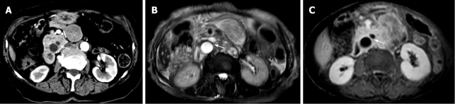
Anomalous junction of the common bile duct and main pancreatic duct. A: CT scan shows the junction of the common bile duct and main pancreatic duct below the level of the uncinate process (arrow); B and C: Axial MR images show the junction of the common bile duct and main pancreatic duct (arrows). (These photographs were processed by Adobe Photoshop Creative Suite 2.).
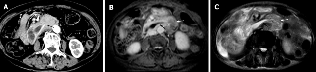
Anomalous termination of the common bile duct. A: Axial image from CT scan shows the termination of the common bile duct at the distal portion of the horizontal part of the duodenum (arrow); B and C: MR images demonstrate a tapered configuration of the common bile duct (black arrows) and a mass located on the proximal jejunum (white arrows). (These photographs were processed by Adobe Photoshop Creative Suite 2.).
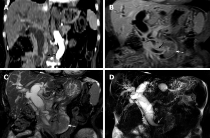
Dilated pancreatic and biliary ducts with ectopic drainage. A: Coronal MDCT image of CT reveals a low termination of the common bile duct (arrow); B and C: Coronal MR images show ectopic pancreaticobiliary junction, low termination of the common bile duct (black arrows), and the mass (white arrows) located on the proximal jejunum; D: MRCP shows dilated pancreatic and biliary ducts. (These photographs were processed by Adobe Photoshop Creative Suite 2.).
Similar articles
-
Sonographic diagnosis of a common pancreaticobiliary channel in children.Pediatr Radiol. 2006 Dec;36(12):1300-5. doi: 10.1007/s00247-006-0322-z. Epub 2006 Oct 7. Pediatr Radiol. 2006. PMID: 17028852
-
A case of anomalous arrangement of the pancreaticobiliary ductal system demonstrated by intraductal ultrasonography.Am J Gastroenterol. 1994 Oct;89(10):1893-5. Am J Gastroenterol. 1994. PMID: 7942691
-
Anomalous pancreaticobiliary duct union associated with bile duct carcinoma.Gastrointest Radiol. 1984;9(1):49-51. doi: 10.1007/BF01887800. Gastrointest Radiol. 1984. PMID: 6724239
-
Pancreaticobiliary maljunction and congenital biliary dilatation.Lancet Gastroenterol Hepatol. 2017 Aug;2(8):610-618. doi: 10.1016/S2468-1253(17)30002-X. Lancet Gastroenterol Hepatol. 2017. PMID: 28691687 Review.
-
Pancreaticobiliary maljunction and carcinogenesis to biliary and pancreatic malignancy.Langenbecks Arch Surg. 2009 Jan;394(1):159-69. doi: 10.1007/s00423-008-0336-0. Epub 2008 May 24. Langenbecks Arch Surg. 2009. PMID: 18500533 Review.
References
-
- Lee HJ, Ha HK, Kim MH, Jeong YK, Kim PN, Lee MG, Kim JS, Suh DJ, Lee SG, Min YI, et al. ERCP and CT findings of ectopic drainage of the common bile duct into the duodenal bulb. AJR Am J Roentgenol. 1997;169:517–520. - PubMed
-
- Rosario MT, Neves CP, Ferreira AF, Luis AS. Ectopic papilla of Vater. Gastrointest Endosc. 1990;36:606–607. - PubMed
-
- Keddie NC, Taylor AW, Sykes PA. The termination of the common bile duct. Br J Surg. 1974;61:623–625. - PubMed
-
- Avisse C, Flament JB, Delattre JF. Ampulla of Vater. Anatomic, embryologic, and surgical aspects. Surg Clin North Am. 2000;80:201–212. - PubMed
Publication types
MeSH terms
LinkOut - more resources
Full Text Sources
Medical
