. 2009 Dec 8;48(48):11421-31.
doi: 10.1021/bi901438z.

Assembly with the Na,K-ATPase alpha(1) subunit is required for export of beta(1) and beta(2) subunits from the endoplasmic reticulum

Affiliations

Assembly with the Na,K-ATPase alpha(1) subunit is required for export of beta(1) and beta(2) subunits from the endoplasmic reticulum

Elmira Tokhtaeva et al. Biochemistry. .

Abstract

The level of the heterodimeric Na,K-ATPase is tightly controlled in epithelia to maintain appropriate transport function. The catalytic Na,K-ATPase alpha subunit is not able to exit the ER or catalyze ion transport unless assembled with the beta subunit. However, requirements for the ER exit of the Na,K-ATPase beta subunit that plays an additional, ion-transport-independent, role in intercellular adhesion are not clear. Exogenous beta(1) or beta(2) subunits expressed in renal MDCK cells replace endogenous beta(1) subunits in the alpha-beta complexes in the ER, resulting in a decrease in the amount of the alpha(1)-bound endogenous beta(1) subunits by 47-61% with no change in the amount of alpha(1) subunits. Disruption of the alpha(1)-beta association by mutations in defined alpha(1)-interacting regions of either beta(1) or beta(2) subunits results in the ER retention and rapid degradation of unassembled mutants. Hence, the ER quality control system allows export only of assembled alpha-beta complexes to the Golgi, thereby maintaining an equimolar ratio of alpha and beta subunits in the plasma membrane, whereas the number of alpha(1) subunits in the ER determines the amount of the alpha-beta complexes.


Figures

FIGURE 1
FIGURE 1
The N-glycans of the immature form of the endogenous Na,K-ATPase β1 subunit are high-mannose type. (A) A simplified scheme showing transformation of high-mannose N-glycans into hybrid- and complex-type N-glycans in the Golgi. The Golgi mannosidases I and II remove terminal mannose residues allowing covalent addition of N-acetylglucosamine residues followed by branching and elongation of N-glycans due to the action of various Golgi glycosyltransferases. A specific inhibitor of the Golgi mannosidase I, deoxymannojirimycin (DMJ), prevents formation of both hybrid- and complex-type N-glycans, thus preserving the high-mannose type structure of N-glycans. A specific inhibitor of the Golgi mannosidase II, swainsonine (Sw), prevents transformation of hybrid-type N-glycans into complex-type N-glycans. Endoglycosidase EndoH can be used as a tool to distinguish the complex-type N-glycans that are resistant to the action of this enzyme from the EndoH-cleavable high-mannose and hybrid-type N-glycans. (B) Confluent monolayers of MDCK cells were incubated in the absence or presence of DMJ (100 µg/mL) or Sw (2 µg/µL) for 48 h and lysed. Cell lysates were analyzed by immunoblotting using the antibodies against the Na, K-ATPase β1 subunit. In the absence of inhibitors, the Na,K-ATPase β1 subunit has two N-glycosylated forms, the mature form (M) and immature form (I). The electrophoretic mobility of the immature form of the Na,K-ATPase β1 subunit is similar to that of the high-mannose form of the β1 subunit formed in the presence of DMJ (left panel, H-M) but greater than that of the hybrid-type β1 subunit formed in the presence of Sw (central panel), indicating that the immature form of the Na,K-ATPase β1 subunit is high-mannose type but not hybrid type. This high-mannose form of the β1 subunit is converted into a deglycosylated product by EndoH (right panel, DG). In contrast, the mature fully glycosylated form of the β1 subunit is resistant to EndoH digestion (right panel), indicating that it contains only complex-type N-glycans.
FIGURE 2
FIGURE 2
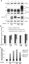
Expression of exogenous Na,K-ATPase β subunits significantly decreases the amount of the complex-type endogenous β1 subunit without a change in the amount of the high-mannose endogenous β1 subunit in MDCK cells. (A) Equal amounts of whole cell lysates of nontransfected cells and cells expressing various exogenous β subunits were analyzed by SDS–PAGE followed by immunoblotting using the antibodies against the α1 and β1 subunits of the Na,K-ATPase, against YFP, and against β-actin. The antibody used for detection of the endogenous canine Na,K-ATPase β1 subunit does not react with rat YFP-β1 but does react with dog YFP-β1. (B, C) Densitometry quantification of the results presented in (A). Quantification of YFP-linked β subunits in cells expressing YFP-β1 (rat), YFP-β2, YFP-β2 (P/G) (lanes 2, 3, and 4 in (A)) relative to the endogenous Na,K-ATPase β1 subunit in nontransfected cells (lane 1 in (A)) was performed by using a conversion factor equal to the ratio between the density of the YFP-β1 (dog) band detected by anti-β1 antibody and the density of the YFP-β1 (dog) band detected by anti-YFP antibody (lane 5 in (A)). (B) Expression of the wild-type YFP-β1 (rat), YFP-β2, and YFP-β1 (dog) decreases the amount of the complex-type glycosylated form of the endogenous Na,K-ATPase β1 subunit by 47–61% but does not alter the amount of the Na,K-ATPase α1 subunit. Expression of the P/G mutant of YFP-β2 does not alter the amount of either the α1 or endogenous β1 subunit. The ratio between the total amount of the Na,K-ATPase β subunit (endogenous and exogenous) and the endogenous α1 subunit in all transfected cell lines is similar to the ratio between β1 and α1 subunits in nontransfected cells. (C) The amount of the high-mannose form of the endogenous β1 subunit is not altered by expression of the exogenous β subunits. The total amount of the high-mannose sub-units (endogenous and exogenous) is significantly increased in all transfected cell lines. Error bars, SD (n = 3). *, significant difference with NT, P < 0.01, Student’s t-test. Key: Na,K-α1 and Na,K-β1, the endogenous α1 and β1 subunits of the Na,K-ATPase; C, complex-type glycosylated form; H, high-mannose glycosylated form.
FIGURE 3
FIGURE 3
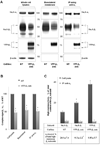
Expression of the exogenous β1 subunit of the Na,K-ATPase decreases the efficiency of the assembly of its high-mannose form with the α1 subunit. (A) The whole cell lysates, basolateral membrane proteins, and proteins immunoprecipitated using the antibody against the Na,K-ATPase α1 subunit were obtained from nontransfected MDCK cells and MDCK cells expressing an YFP-linked rat β1 subunit as described in Materials and Methods. All three protein fractions were analyzed by SDS–PAGE followed by immunoblotting using the antibodies against the Na,K-ATPase α1 subunit, Na,K-ATPase β1 subunit, YFP, and β-actin. The antibody used for detection of the endogenous canine Na,K-ATPase β1 subunit does not react with the rat β1 subunit of YFP-β1. The β-actin was used as a loading control in whole cell lysates and as a negative control in basolateral and immunoprecipitated fractions to show no contamination by the nonrelated proteins of the cell lysate. (B, C) Densitometry quantification of the results presented in (A). (B) Expression of YFP-β1 decreases the ratio between the amount of the complex-type glycosylated form of the endogenous β1 subunit and the amount of the α1 subunit in whole cell lysates, basolateral membrane fractions, and α1-immunoprecipitated fractions by 46–52%. (C) The percentages of the high-mannose forms of both the endogenous Na,K-ATPase β1 subunit and YFP-β1 are higher in whole cell lysates than in α1-immunoprecipitates, showing that only some of the high-mannose subunits are assembled with the α1 subunit. The fraction of the α1-bound high-mannose β1 subunits of total high-mannose β1 subunits that was calculated as the ratio between percentages of the high-mannose β1 subunit in α1-immunoprecipitated fractions and whole cell lysates is decreased by expression of YFP-β1 (see table). Error bars and errors, SD (n = 3). *, significant difference with NT, P < 0.01, Student’s t-test. Key: Na,K-α1 and Na,K-β1, the endogenous α1 and β1 subunits of the Na,K-ATPase; C, complex-type glycosylated form; H, high-mannose glycosylated form.
FIGURE 4
FIGURE 4
High-mannose forms of the endogenous β1 subunit and YFP-β1 are degraded more rapidly than the complex-type subunits. (A) Tight monolayers of nontransfected and YFP-β1-expressing MDCK cells were incubated in the presence of 20 µg/mL cycloheximide (CHX) for the indicated time periods and lysed. Equal volumes of the whole cell lysates were analyzed by Western blot using the antibodies against the Na,K-ATPase β1 subunit and against YFP. The level of high-mannose forms of both Na,K-ATPase β1 subunit and YFP-β1 decreases much more rapidly than the amount of the complex-type forms during 5 h incubation with CHX. (B) Densitometry of the results presented in (A) shows that the amount of complex-type β1 subunit decreases only by 10% after 5 h incubation with CHX. In contrast, the amount of the high-mannose β1 subunit decreases by 50–60% after only 1 h incubation. (C) The Na,K-ATPase α1 subunit was immunoprecipitated from lysates of nontransfected and YFP-β1-expressing MDCK cells incubated in the absence or presence of CHX for 1 h as described in Materials and Methods. Coprecipitated Na,K-ATPase β1 subunits were analyzed by Western blot. In the absence of the inhibitor, the α1-bound β1 subunits contain a predominant fraction of complex-type and a minor fraction of the high-mannose-type glycoforms in both nontransfected and transfected cells. The high-mannose fraction is not detected after 1 h incubation with CHX in either cell line even after overexposure of the blots (lanes 2). Error bars and errors, SD (n = 3). Key: Na,K-β1, the endogenous β1 subunit of the Na, K-ATPase; C, complex-type glycosylated form; H, high-mannose glycosylated form.
FIGURE 5
FIGURE 5
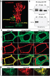
Impairment of α–β association due to the point mutations in the α-interacting regions of the β2 subunit correlates with retention of the mutants in the ER. (A) A model of the Na,K-ATPase α1 and β1 subunits based on the crystal structure of the sodium–potassium pump at 2.4 Å resolution (2ZXE) (13) shows positions of a triple mutation, Y39A/F42A/Y43A (YFY/AAA), and a single mutation, P245G (P/G), that were introduced into putative α1-interacting regions of the YFP-linked rat β1 subunit. The model shows that Y39, F42, and Y43 are located at the interface between the transmembrane domain of the β1 subunit and the seventh transmembrane domain (TM7) of the α1 subunit and P245 is located at the interface between the extracellular domain of the β1 subunit and the 7–8 loop of the α1 subunit. Numbering of the amino acid residues corresponds to the rat β1 subunit. The residues homologous to Y39, F42, Y43A, and P245were also mutated in the YFP-linked human β2 subunit based on the alignment shown in Supporting Information Figure 1, producing a triple mutant, Y44A/F47A/Y48A (YFY/AAA), and a single mutant, P229G (P/G), of YFP-β2. (B) The wild-type YFP-β2 and its YFY/AAA and P/G mutants were immunoprecipitated from the respective cell lysates using the antibody against YFP. Precipitated YFP-linked proteins and coprecipitated endogenous α1 subunit were analyzed by immunoblotting. The percentage of the high-mannose form of YFP-β2 is increased in the mutants (top panel). Coimmunoprecipitation of the α1 subunit with YFP-β2 is decreased by YFY/AAA mutations and abolished by P/G mutation (bottom panel). (C) Immunostaining of the endogenous α1 subunit shows that the wild-type YFP-β2 precisely colocalizes, the YFY/AAA mutant only partially colocalizes, and the P/G mutant does not colocalize with the α1 subunit in the basolateral membrane. (D) Transient transfection of cells expressing the P/G mutant of YFP-β2 with the fluorescent marker of the ER, DsRed2-ER, shows colocalization of the mutant and the ER. Key: Na,K-α1, the endogenous α1 subunit of the Na,K-ATPase; C, complex-type glycosylated form; H, high-mannose glycosylated form.

References

    1. Xie Z. Molecular mechanisms of Na/K-ATPase-mediated signal transduction. Ann. N.Y. Acad. Sci. 2003;986:497–503. - PubMed
    1. Tian J, Xie ZJ. The Na-K-ATPase and calcium-signaling microdomains. Physiology (Bethesda, MD) 2008;23:205–211. - PMC - PubMed
    1. Blanco G, Mercer RW. Isozymes of the Na-K-ATPase: heterogeneity in structure, diversity in function. Am. J. Physiol. 1998;275:F633–F650. - PubMed
    1. Crambert G, Hasler U, Beggah AT, Yu C, Modyanov NN, Horisberger JD, Lelievre L, Geering K. Transport and pharmacological properties of nine different human Na, K-ATPase isozymes. J. Biol. Chem. 2000;275:1976–1986. - PubMed
    1. Sweadner KJ, Rael E. The FXYD gene family of small ion transport regulators or channels: cDNA sequence, protein signature sequence, and expression. Genomics. 2000;68:41–56. - PubMed

Publication types

Substances