

Irinotecan-induced mucositis manifesting as diarrhoea corresponds with an amended intestinal flora and mucin profile
- PMID: 19765103
- PMCID: PMC2768147
- DOI: 10.1111/j.1365-2613.2009.00671.x
Irinotecan-induced mucositis manifesting as diarrhoea corresponds with an amended intestinal flora and mucin profile
Abstract
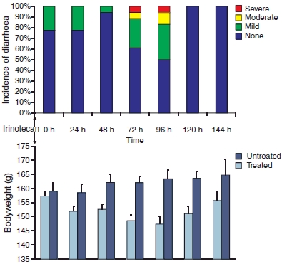
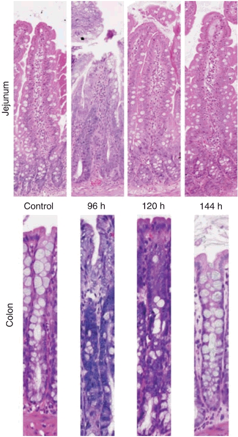
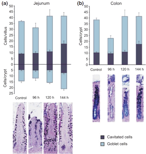
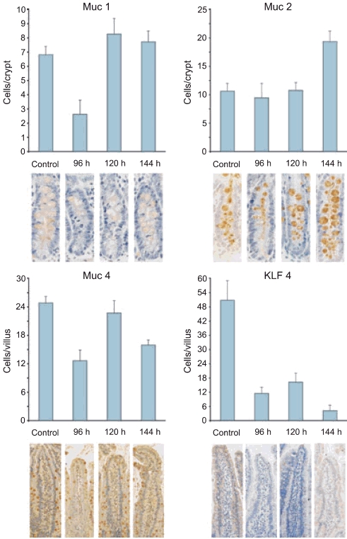
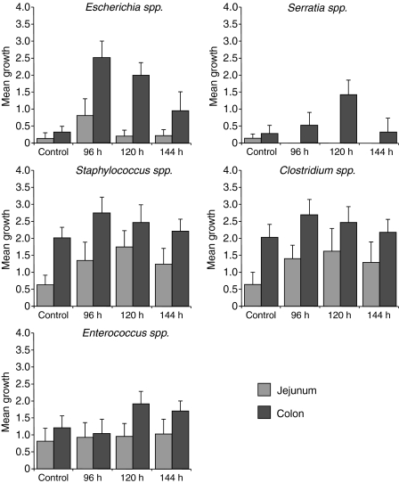
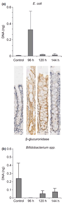
Chemotherapy-induced diarrhoea is a major oncological problem, caused by the cytotoxic effects of cancer chemotherapy. Irinotecan is linked with severe mucositis and diarrhoea, the mechanisms of which remain poorly understood. Bacterial beta-glucuronidase is thought to be involved in the metabolism of irinotecan, implicating the intestinal flora. Intestinal mucins may also be implicated in the development of chemotherapy-induced diarrhoea. Rats were treated with 200 mg/kg of irinotecan and killed at 96, 120 and 144 h. The rats were monitored for diarrhoea. Pathology and immunohistochemical staining was performed. The samples were cultured and faecal DNA was analysed using real-time polymerase chain reaction. Severe diarrhoea was observed from 72 to 96 h. A decrease in body mass was also observed after treatment. Significant changes in goblet cell numbers (both complete and cavitated cells) were observed in the small and large intestines. Changes in MUC gene expression were observed in the small intestine only. Modifications were observed to the intestinal flora profile, especially Escherichia coli, and an increase in the expression of beta-glucuronidase was detected. In conclusion, irinotecan-induced diarrhoea may be caused by an increase in some beta-glucuronidase-producing bacteria, especially E. coli, exacerbating the toxicity of active metabolites. Accelerated mucous secretion and mucin release may also contribute to the delayed onset of diarrhoea.
Figures
Similar articles
-
Irinotecan-induced mucositis is associated with changes in intestinal mucins.Cancer Chemother Pharmacol. 2009 Jun;64(1):123-32. doi: 10.1007/s00280-008-0855-y. Epub 2008 Nov 8. Cancer Chemother Pharmacol. 2009. PMID: 18998135
-
Characterisation of mucosal changes in the alimentary tract following administration of irinotecan: implications for the pathobiology of mucositis.Cancer Chemother Pharmacol. 2008 Jun;62(1):33-41. doi: 10.1007/s00280-007-0570-0. Epub 2007 Aug 17. Cancer Chemother Pharmacol. 2008. PMID: 17703303
-
Irinotecan induces enterocyte cell death and changes to muc2 and muc4 composition during mucositis in a tumour-bearing DA rat model.Cancer Chemother Pharmacol. 2019 May;83(5):893-904. doi: 10.1007/s00280-019-03787-5. Epub 2019 Feb 27. Cancer Chemother Pharmacol. 2019. PMID: 30815720
-
Irinotecan- and 5-fluorouracil-induced intestinal mucositis: insights into pathogenesis and therapeutic perspectives.Cancer Chemother Pharmacol. 2016 Nov;78(5):881-893. doi: 10.1007/s00280-016-3139-y. Epub 2016 Sep 2. Cancer Chemother Pharmacol. 2016. PMID: 27590709 Review.
-
Chemotherapy-induced mucositis: the role of gastrointestinal microflora and mucins in the luminal environment.J Support Oncol. 2007 Jun;5(6):259-67. J Support Oncol. 2007. PMID: 17624050 Review.
Cited by
-
Low-dose irradiation affects the functional behavior of oral microbiota in the context of mucositis.Exp Biol Med (Maywood). 2016 Jan;241(1):60-70. doi: 10.1177/1535370215595467. Epub 2015 Jul 22. Exp Biol Med (Maywood). 2016. PMID: 26202372 Free PMC article.
-
The role of intestinal microbiota in the development and severity of chemotherapy-induced mucositis.PLoS Pathog. 2010 May 27;6(5):e1000879. doi: 10.1371/journal.ppat.1000879. PLoS Pathog. 2010. PMID: 20523891 Free PMC article. Review.
-
Current evidence for vitamin D in intestinal function and disease.Exp Biol Med (Maywood). 2019 Sep;244(12):1040-1052. doi: 10.1177/1535370219867262. Epub 2019 Jul 31. Exp Biol Med (Maywood). 2019. PMID: 31366237 Free PMC article. Review.
-
Managing Irinotecan-Induced Diarrhea: A Comprehensive Review of Therapeutic Interventions in Cancer Treatment.Pharmaceuticals (Basel). 2025 Mar 2;18(3):359. doi: 10.3390/ph18030359. Pharmaceuticals (Basel). 2025. PMID: 40143136 Free PMC article. Review.
-
Oral mucositis: the new paradigms.Curr Opin Oncol. 2010 Jul;22(4):318-22. doi: 10.1097/CCO.0b013e32833a9fab. Curr Opin Oncol. 2010. PMID: 20485169 Free PMC article. Review.
References
-
- Araki E, Ishikawa M, Iigo M, Koide T, Itabashi M, Hoshi A. Relationship between development of diarrhea and the concentration of SN-38, an active metabolite of CPT-11, in the intestine and the blood plasma of athymic mice following intraperitoneal administration of CPT-11. Jpn. J. Cancer Res. 1993;84:697–702. - PMC - PubMed
-
- Armand JP, Ducreux M, Mahjoubi M, et al. CPT-11 (irinotecan) in the treatment of colorectal cancer. Eur. J. Cancer. 1995;31A:1283–1287. - PubMed
-
- Bowen JM, Stringer AM, Gibson RJ, Yeoh AS, Hannam S, Keefe DM. VSL#3 probiotic treatment reduces chemotherapy-induced diarrhea and weight loss. Cancer Biol. Ther. 2007;6:1449–1454. - PubMed
-
- Brandi G, Dabard J, Raibaud P, et al. Intestinal microflora and digestive toxicity of irinotecan in mice. Clin. Cancer Res. 2006;12:1299–1307. - PubMed
Publication types
MeSH terms
Substances
LinkOut - more resources
Full Text Sources
Medical
