. 2010 Jun;14(6B):1453-67.
doi: 10.1111/j.1582-4934.2009.00909.x. Epub 2009 Sep 18.

Anti-CD44 induces apoptosis in T lymphoma via mitochondrial depolarization

Affiliations

Anti-CD44 induces apoptosis in T lymphoma via mitochondrial depolarization

Mohini Rajasagi et al. J Cell Mol Med. 2010 Jun.

Abstract

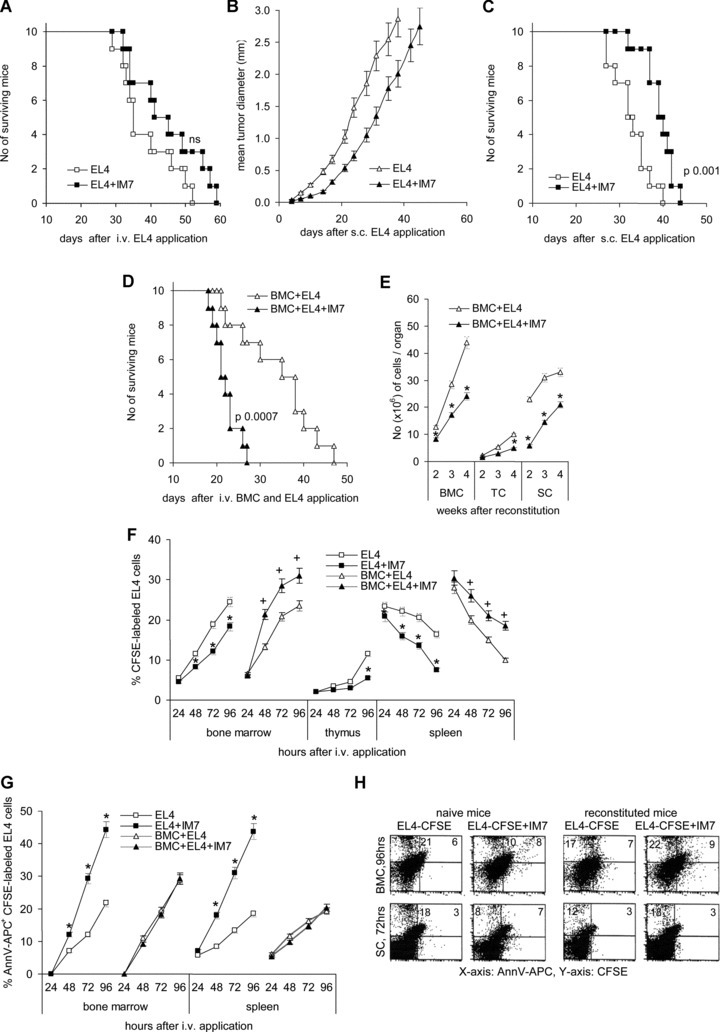
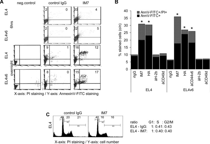
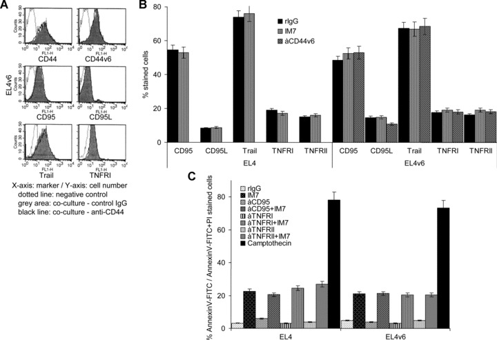
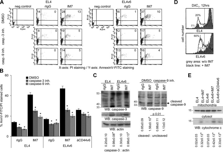
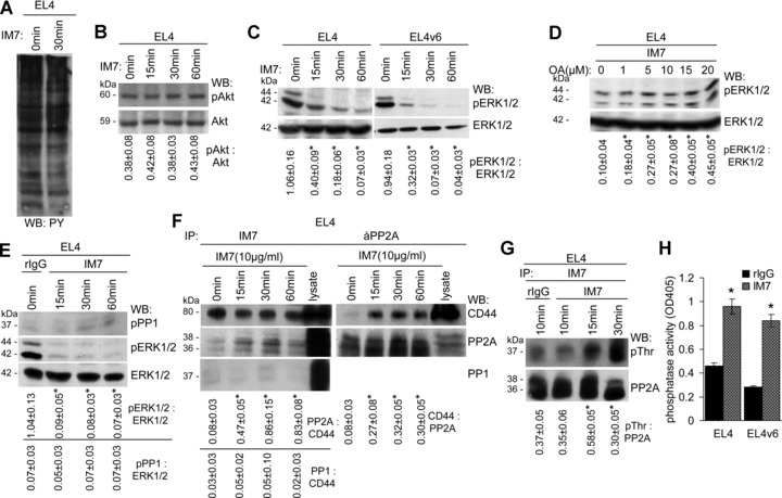
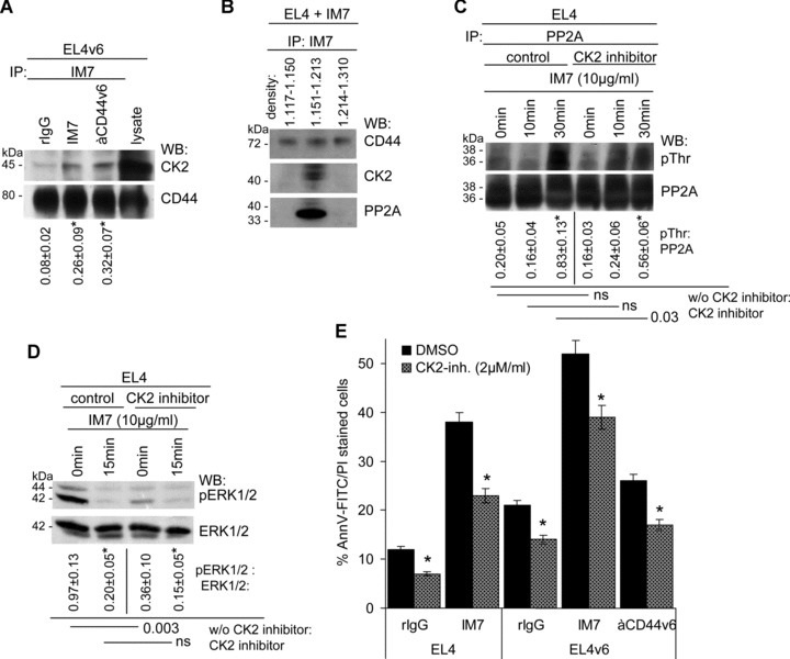
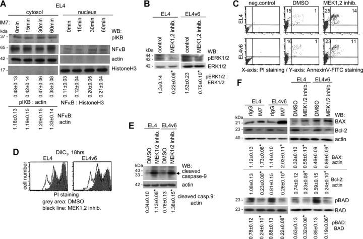
A blockade of CD44 can interfere with haematopoietic and leukemic stem cell homing, the latter being considered as a therapeutic option in haematological malignancies. We here aimed to explore the molecular mechanism underlying the therapeutic efficacy of anti-CD44. We noted that in irradiated mice reconstituted with a bone marrow cell transplant, anti-CD44 exerts a stronger effect on haematopoietic reconstitution than on T lymphoma (EL4) growth. Nonetheless, in the non-reconstituted mouse anti-CD44 suffices for a prolonged survival of EL4-bearing mice, where anti-CD44-prohibited homing actively drives EL4 cells into apoptosis. In vitro, a CD44 occupancy results in a 2-4-fold increase in apoptotic EL4 cells. Death receptor expression (CD95, TRAIL, TNFRI) remains unaltered and CD95 cross-linking-mediated apoptosis is not affected. Instead, CD44 ligation promotes mitochondrial depolarization that is accompanied by caspase-9 cleavage and is inhibited in the presence of a caspase-9 inhibitor. Apoptosis becomes initiated by activation of CD44-associated phosphatase 2A (PP2A) and proceeds via ERK1/2 dephosphorylation without ERK1/2 degradation. Accordingly, CD44-induced apoptosis could be mimicked by ERK1/2 inhibition, that also promotes EL4 cell apoptosis through the mitochondrial pathway. Thus, during haematopoietic stem cell reconstitution care should be taken not to interfere by a blockade of CD44 with haematopoiesis, which could be circumvented by selectively targeting leukemic CD44 isoforms. Beyond homing/settlement in the bone marrow niche, anti-CD44 drives leukemic T cells into apoptosis via the mitochondrial death pathway by CD44 associating with PP2A. Uncovering this new pathway of CD44-induced leukemic cell death provides new options of therapeutic interference.


Figures

Fig 1
Fig 1
The impact of anti-CD44 on thymoma growth in vivo: (A–E) C57BL6 mice received an i.v. (A) or s.c. (B, C) injection of 104 EL4 cells or (C, D) were lethally irradiated and reconstituted with 1 × 10⁷ T-cell-depleted BMC and received EL4 cells, i.v. 2 days after reconstitution. Mice received twice per week an i.v. injection of 150 μg control IgG or IM7. (A) The survival time of mice receiving EL4 cells i.v. is shown. Survival was slightly, but not to a significant level prolonged by IM7 application. (B, C) After s.c. application of EL4 cells, IM7 retarded the start of the tumour growth and the survival time became significantly prolonged. (D) The survival time of IM7-treated, EL4 tumour-bearing reconstituted mice was significantly shortened as compared to mice receiving control IgG and (E) the recovery of BMC, TC and SC (mean number ± S.D. of three mice/group) was significantly delayed (*). (F) C57BL6 mice were treated as described above, but received 2 × 10⁶ CFSE-labelled EL4 cells. The% of CFSE-labelled EL4 cells in bone marrow, thymus and spleen was evaluated during 96 hrs. Mean values ± S.D. of three mice are shown. The recovery of tumour cells is significantly reduced in IM7-treated, non-reconstituted mice (*), but unaltered or increased in IM7-treated reconstituted mice (+). (G) The percentage (mean ± S.D.) of apoptotic CFSE-labelled EL4 cells during the starting 96 hrs after i.v. application was evaluated by annexinV-APC staining. The percentage of apoptotic EL4 cells is significantly increased in non-reconstituted IM7-treated mice (*), but largely unaltered in reconstituted IM7-treated mice. (H) Examples of apoptotic EL4 cells in the bone marrow 96 hrs and in the spleen 72 hrs after application of CFSE-labelled EL4 cells in naive and reconstituted C57BL6 mice.
Fig 2
Fig 2
CD44 ligation induces apoptosis in EL4 cells: (A and B) EL4 and EL4v6 cells were cultured for 6 hrs or o/n in the presence of 10 μg/ml IM7, anti-CD44v6 or rIgG or 15 μg/ml HA and, as controls, 10 μg/ml anti-H-2b or anti-CD49d. Cells were stained with annexinV-FITC/PI. A representative example (A) and mean values ± S.D. of apoptotic cells are shown (B). Significant differences in comparison to cells cultured in the presence of rIgG are indicated by *. (C) Cells were stained with PI and cultured o/n in the presence of rIgG or IM7. The percentage of cells in G1, S and G2 or M phase was evaluated by flow cytometry. Cell cycle progression did not vary significantly in dependence on the presence of IM7.
Fig 3
Fig 3
EL4 and EL4v6 cells are resistant towards Fas-induced apoptosis: (A and B) CD44, CD44v6, CD95, CD95L, Trail, TNFRI and TNFRII expression on EL4 cells and EL4v6 cells was evaluated after o/n incubation in the presence of rIgG, IM7 or anti-CD44v6 (10 μg/ml). (A) A representative example and (B) mean values ± S.D. are shown. IM7 had no impact on CD44 and apoptosis receptor expression. (C) EL4 and EL4v6 cells were cultured o/n on uncoated or anti-CD95-, anti-TNFRI- or anti-TNFRII-coated plates. Cultures contained 10 μg/ml rIgG or IM7 or 1 μM/ml camptothecin (positive control). The percentage (mean ± S.D. of triplicates) of annexinV-FITC/PI stained cells is shown. EL4 and EL4v6 cells were resistant towards receptor-induced apoptosis and IM7-induced apoptosis was independent of death receptor cross-linking.
Fig 4
Fig 4
CD44 ligation induces apoptosis via the mitochondrial pathway: (A and B) EL4 and EL4v6 cells were cultured o/n in the presence of rIgG, IM7 or anti-CD44v6. Cultures contained in addition DMSO (control) or 2 μM caspase-3 or caspase-9 inhibitor. Apoptosis was measured by annexinV-FITC/PI staining. (A) A representative example and (B) mean values ± S.D. of triplicates are shown. Significant differences in the percentage of apoptotic cells in the presence of caspase inhibitors are indicated by *. (C) WB of caspase-3 and caspase-9 cleavage in lysates of EL4 cells cultured o/n in the presence of rIgG or IM7 or in the presence of a caspase-9 inhibitor. The mean values ± S.D. of the ratio of caspase-3: actin and cleaved: uncleaved caspase-9 are shown. Significant differences by IM7 or a caspase-9 inhibitor are indicated by an asterisk. Caspase-3 and capase-9 cleavage is enhanced in IM7 treated cells and caspase-9 cleavage is reduced in the presence of the caspase-9 inhibitor. (D) EL4 cells were stained with the mitochondrial dye DilC1 and incubated with 10 μg/ml IM7 for 12 hrs. DilC1 staining was evaluated by flow cytometry. (E) EL4 cells were incubated with 10 μg/ml IM7 for 12 hrs. Mitochondria were separated from the cytosol. Lysates were separated by SDS-PAGE, proteins were transferred to a nitrocellulose membrane and blotted with anti-cytochrome c. The mean values ± S.D. of the ratio of cytochrome c in the cytosol: mitochondria is shown. Significant differences by IM7 or anti-CD44v6 are indicated by an asterisk. (D and E) Mitochondria integrity is strongly affected after 12 hrs of culture in the presence of IM7.
Fig 5
Fig 5
CD44 ligation induces PP2A activation and ERK1/2 dephosphorylation: (A) EL4 cells were cultured for 30 min. in the presence of IM7. Cells were lysed and proteins were separated by SDS-PAGE, transferred to a nitrocellulose membrane and probed with anti-phosphotyrosine. (B and C) EL4 and EL4v6 cells were cultured for 15–60 min. in the presence of IM7. After lysis, SDS-PAGE and transfer, membranes were incubated with (B) anti-Akt and anti-pAkt and (C) anti-ERK1/2 and anti-pERK1/2. The mean values ± S.D. of the ratio of pAkt: Akt and pERK1/2: ERK1/2 are shown. Significant differences by IM7 are indicated by an asterisk. Tyrosine and Akt phosphorylation remained unaltered. ERK1/2 phosphorylation of EL4 and EL4v6 cells cultured in the presence of IM7 becomes strikingly reduced. (D) EL4 cells were cultured in the presence of rIgG or IM7 and increasing amounts of OA. Cells were lysed, lysates were separated by SDS-PAGE, transferred and blotted with anti-ERK1/2 and anti-pERK1/2. The mean values ± S.D. of the ratio of pERK1/2: ERK1/2 are shown. Significant differences by OA are indicated by an asterisk. In the presence of the phosphatase inhibitor, the IM7-induced reduction of ERK1/2 phosphorylation was prevented. (E) EL4 cells were cultured in the presence of rIgG or IM7. Cells were lysed, lysates were separated by SDS-PAGE, transferred and blotted with anti-pPP1, anti-ERK1/2 and anti-pERK1/2. The mean values ± S.D. of the ratio of pPP1: ERK1/2 and for comparison of pERK1/2: ERK1/2 are shown. Significant differences by IM7 are indicated by an asterisk. PP1 did not become phosphorylated concomitantly with ERK1/2 dephosphorylation. (F) EL4 cells were cultured in the presence of IM7. Cells were lysed and immunoprecipitated with IM7 or anti-PP2A. Precipitates were separated by SDS-PAGE, transferred and blotted with IM7, anti-PP2A and anti-PP1. The mean values ± S.D. of the ratio of PP2A: CD44, PP1: CD44, respectively, of CD44: PP2A are shown. Significant differences by IM7 are indicated by an asterisk. PP2A, but not PP1 co-immunoprecipitated with CD44 and vice versa. (G) EL4 cells were cultured in the presence of rIgG or IM7. Cells were lysed and lysates were immunoprecipitated with IM7. Lysates were separated by SDS-PAGE, transferred and blotted with anti-PP2A and anti-pThr. The mean values ± S.D. of the ratio of pThr: PP2A are shown. Significant differences by IM7 are indicated by an asterisk. Threonine phosphorylation of PP2A became strengthened in the presence of IM7. (H) Phosphatase activity of the anti-CD44 immunoprecipitate was tested by ELISA. Phosphatase activity was strongly increased after culture of EL4 and EL4v6 cells in the presence of IM7.
Fig 6
Fig 6
CK2 is associated with CD44 and promotes partial PP2A activation: (A) EL4 cells were cultured in the presence of rIgG or IM7. Cells were lysed and lysates were immunoprecipitated with IM7. After SDS-PAGE of the precipitate, proteins were transferred and blotted with anti-CD44 and anti-CK2. The mean values ± S.D. of the ratio of CK2: CD44 are shown. Significant differences by IM7 and anti-CD44v6 are indicated by an asterisk. The CD44 precipitate contained low amounts of CK2. (B) EL4 cells, cultured in the presence of IM7, were lysed and light and dense membrane fractions were separated by sucrose gradient centrifugation. Light, medium dense and heavy fractions were pooled and immunoprecipitated with IM7. After SDS-PAGE and transfer, membranes were blotted with IM7, anti-PP2A and anti-CK2. PP2A and CK2 co-immunoprecipitated with CD44 in the pooled fractions of density 1.15–1.21. (C) EL4 cells were cultured in the presence of IM7 and, where indicated a CK2 inhibitor (2 μM/ml). Cells were lysed and the lysates were precipitated with anti-PP2A, separated by SDS-PAGE, transferred and blotted with anti-PP2A and anti-pThr. The mean values ± S.D. of the ratio of pThr: PP2A are shown. Significant differences by IM7 and the CK2 inhibitor are indicated. The CK2 inhibitor partly inhibited PP2A phosphorylation. (D) EL4 cells were cultured in the presence of IM7 and, where indicated the CK2 inhibitor, lysates were precipitated by SDS-PAGE, transferred and blotted with anti-ERK1/2 and anti-pERK1/2. The mean values ± S.D. of the ratio of pERK1/2: ERK1/2 are shown. Significant differences by IM7 and the CK2 inhibitor are indicated. ERK1/2 phosphorylation was reduced in the presence of the CK2 inhibitor. (E) EL4 and EL4v6 cells were cultured in the presence of rIgG, IM7 or anti-CD44v6. Where indicated the cultures contained a CK2 inhibitor. Apoptosis was evaluated after 24 hrs by annexinV-FITC/PI staining. The CK2 inhibitor interfered with apoptosis induction independent of the presence of IM7 or anti-CD44v6.
Fig 7
Fig 7
ERK1/2 dephosphorylation promotes mitochondrial membrane disintegration and caspase-9 cleavage: (A) EL4 cells were cultured for 15–60 min. in the presence of IM7. After separation of the cytosolic from the nuclear fraction, lysates were separated by SDS-PAGE, transferred and blotted with anti-pIKB and anti-NFκB. Actin and HistoneH3 served as controls. The mean values ± S.D. of the ratio of pIKB and NFκB: actin (cytosol) and of NF-kB: HistoneH3 (nucleus) are shown. Significant differences by IM7 are indicated by an asterisk. IM7 treatment did not influence IKB phosphorylation and did not significantly alter NFκB liberation. (B–F) EL4 and EL4v6 cells were cultured o/n in the presence of a MEK1,2 inhibitor. (B) The mean values ± S.D. of the ratio of pERK1/2: ERK1/2 are shown. Significant differences by the MEK1,2 inhibitor are indicated by an asterisk. ERK1/2 phosphorylation in EL4 and EL4v6 cells was inhibited in the presence of MEK1,2 inhibitor. (C) Apoptosis (annexinV-FITC/PI staining) was significantly increased in the presence of the MEK1,2 inhibitor. (D) Cells were stained with DilC1 and mitochondrial membrane integrity was determined after 18 hrs incubation by flow cytometry. In the presence of the MEK1,2 inhibitor, mitochondrial membrane integrity was decreased. (E) Cells were lysed, proteins were separated by SDS-PAGE, transferred and blotted with anti-caspase-9. The mean values ± S.D. of the ratio of cleaved caspase-9: actin are shown. Significant differences by IM7 and the MEK1,2 inhibitor are indicated by an asterisk. Caspase-9 cleavage was pronounced in the presence of the MEK1,2 inhibitor. (F) Cells were cultured o/n in the presence of the MEK1,2 inhibitor or in the presence of IM7. Cells were lysed and proteins were separated by SDS-PAGE, transferred and blotted with anti-BAX, anti-Bcl-2, anti-BAD and anti-pBAD. The mean values ± S.D. of the ratio of BAX and Bcl-2: actin and of pBAD: BAD are shown. Significant differences by IM7 and the MEK1,2 inhibitor are indicated by an asterisk. The MEK1,2 inhibitor and IM7 promoted BAX activation and prohibited Bcl-2 expression and BAD phosphorylation.

Similar articles

Cited by

References

    1. Screaton GR, Bell MV, Jackson DG, et al. Genomic structure of DNA encoding the lymphocyte homing receptor CD44 reveals at least 12 alternatively spliced exons. Proc Natl Acad Sci USA. 1992;89:12160–4. - PMC - PubMed
    1. Lynch KW. Consequences of regulated pre-mRNA splicing in the immune system. Nat Rev Immunol. 2004;4:931–40. - PubMed
    1. Naor D, Wallach-Dayan SB, Zahalka MA, Sionov RV. Involvement of CD44, a molecule with a thousand faces, in cancer dissemination. Semin Cancer Biol. 2008;18:260–7. - PubMed
    1. Orian-Rousseau V, Ponta H. Adhesion proteins meet receptors: a common theme. Adv Cancer Res. 2008;101:63–92. - PubMed
    1. Lapidot T, Dar A, Kollet O. How do stem cells find their way home? Blood. 2005;106:1901–10. - PubMed

Publication types

MeSH terms