. 2009;11(5):R138.
doi: 10.1186/ar2810. Epub 2009 Sep 18.

Suppressive effect of secretory phospholipase A2 inhibitory peptide on interleukin-1beta-induced matrix metalloproteinase production in rheumatoid synovial fibroblasts, and its antiarthritic activity in hTNFtg mice

Affiliations

Suppressive effect of secretory phospholipase A2 inhibitory peptide on interleukin-1beta-induced matrix metalloproteinase production in rheumatoid synovial fibroblasts, and its antiarthritic activity in hTNFtg mice

Maung-Maung Thwin et al. Arthritis Res Ther. 2009.

Abstract

Introduction: Secretory phospholipase A2 (sPLA2) and matrix metalloproteinase (MMP) inhibitors are potent modulators of inflammation with therapeutic potential, but have limited efficacy in rheumatoid arthritis (RA). The objective of this study was to understand the inhibitory mechanism of phospholipase inhibitor from python (PIP)-18 peptide in cultured synovial fibroblasts (SF), and to evaluate its therapeutic potential in a human tumor necrosis factor (hTNF)-driven transgenic mouse (Tg197) model of arthritis.

Methods: Gene and protein expression of sPLA2-IIA, MMP-1, MMP-2, MMP-3, MMP-9, tissue inhibitor of metalloproteinase (TIMP)-1, and TIMP-2 were analyzed by real time PCR and ELISA respectively, in interleukin (IL)-1beta stimulated rheumatoid arthritis (RA) and osteoarthritis (OA) synovial fibroblasts cells treated with or without inhibitors of sPLA2 (PIP-18, LY315920) or MMPs (MMP Inhibitor II). Phosphorylation status of mitogen-activated protein kinase (MAPK) proteins was examined by cell-based ELISA. The effect of PIP-18 was compared with that of celecoxib, methotrexate, infliximab and antiflamin-2 in Tg197 mice after ip administration (thrice weekly for 5 weeks) at two doses (10, 30 mg/kg), and histologic analysis of ankle joints. Serum sPLA2 and cytokines (tumor necrosis factor (TNF)alpha, IL-6) were measured by Escherichia coli (E coli) assay and ELISA, respectively.

Results: PIP-18 inhibited sPLA2-IIA production and enzymatic activity, and suppressed production of MMPs in IL-1beta-induced RA and OA SF cells. Treatment with PIP-18 blocked IL-1beta-induced p38 MAPK phosphorylation and resulted in attenuation of sPLA2-IIA and MMP mRNA transcription in RA SF cells. The disease modifying effect of PIP-18 was evidenced by significant abrogation of synovitis, cartilage degradation and bone erosion in hTNF Tg197 mice.

Conclusions: Our results demonstrate the benefit that can be gained from using sPLA2 inhibitory peptide for RA treatment, and validate PIP-18 as a potential therapeutic in a clinically relevant animal model of human arthritis.


Figures
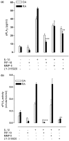
Figure 1
Figure 1
Inhibition of sPLA2-IIA release into medium by PIP-18 in RA and OA SF cultures. Confluent synovial fibroblast (SF) cells in 75 cm2 flasks were serum-starved for overnight (16 hours) before incubation for one hour with 5 μM PIP-18, LY315920, matrix metalloproteinase inhibitor II (MMP-II), or with vehicle (0.5% dimethyl sulfoxide final concentration in medium), and stimulation with hrIL-1β (10 ng/ml) for 24 hours. Rheumatoid arthritis (RA)/osteoarthritis (OA) SFs cultured without IL-1β or the inhibitors served as controls. (a) Immunoreactive secretory phospholipase A2 (sPLA2) released in the culture medium was determined by sPLA2 human type IIA enzyme-linked immunoassay kit. (b) sPLA2 enzymatic activity was measured with an Escherichia coli membrane assay as described [11]. Data shown are the mean ± standard error of the mean of the combined data of triplicate determination of triplicate experiments performed on a pool of RA SF cultures from five RA patients. One-way analysis of variance with post hoc test was done using Bonferroni's correction. *P < 0.05, **P < 0.001, ***P < 0.001 for pair-wise comparisons of each inhibitor type (IL without inhibitor versus IL with inhibitor). PIP = phospholipase inhibitor from python.
Figure 2
Figure 2
Suppressive effects of PIP-18 versus sPLA2 and MMP inhibitors on MMP secretion. Osteoarthritis (OA) and rheumatoid arthritis (RA) synovial fibroblast (SF) cells were incubated for one hour with 5 μM phospholipase inhibitor from python (PIP)-18, matrix metalloproteinase (MMP)-II inhibitor or secretory phospholipase A2(sPLA2) inhibitor LY-315920, stimulated overnight with rhIL-1β (10 ng/ml), and supernatants assayed for MMP secretions by ELISA: (a) MMP-1, (b) MMP-3, (c) MMP-2, (d) MMP-9, (e) tissue inhibitor of metalloproteinase (TIMP)-1, (f) TIMP-2. Results are the mean ± standard error of the mean of the combined data of triplicate determination of triplicate experiments done on a pool of RA SF cultures from five RA patients. Bonferroni's post hoc test was done only if the analysis of variance single-factor test was found significant. *P < 0.05, **P < 0.01, ***P < 0.001 for pair-wise comparisons (IL without inhibitor versus IL with each of the inhibitor used in the study.
Figure 3
Figure 3
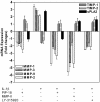
Peptide treatment inhibited MMP and sPLA2 gene expression in IL-1β induced RA SF. Cells were pretreated with the peptide (phospholipase inhibitor from python (PIP)-18), secretory phospholipase A2 (sPLA2) inhibitor (LY315920) or matrix metalloproteinase inhibitor (MMP-II) at 5 μM for one hour, and incubated with hrIL-1β (10 ng/ml) for 24 hours before isolating total RNA. Relative mRNA expression levels were determined by real-time PCR analyses, normalized to internal GAPD values, and plotted relative to control samples treated with vehicle (0.5% dimethyl sulfoxide). Gene-specific real-time analysis was performed for all seven mRNA targets, sPLA2, MMP-1, -2, -3, -9, tissue inhibitor of metalloproteinase (TIMP)-1 and TIMP-2. Results shown are the mean ± standard deviation of fold inductions from three independent experiments with a pool of rheumatoid arthritis (RA) synovial fibroblast (SF) cultures obtained from five RA patients.
Figure 4
Figure 4
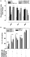
PIP-18 suppresses IL-stimulated p38 MAPK phosphorylation. (a) Rheumatoid arthritis (RA) synovial fibroblast (SF) cells were preincubated at 37°C for one hour with various inhibitors at optimal concentrations: phospholipase inhibitor from python (PIP)-18 (5 μM), LY315920 (5 μM), SB202190 (10 μM), PD98059 (1 μM) or SP600125 (5 μM), and stimulated with rhIL-1β (10 ng/ml) for 30 minutes before assaying for p38, Erk and JNK phosphorylation, using cell-based ELISA. For control of systematic variation, blank control wells (without cells) as well as experimental control wells (seeded cells without any treatment) were included. Phosphorylation index (Pi) was calculated as relative levels of the phosphorylated form of mitogen-activated protein kinase (MAPK)/total MAPK levels. Values are mean ± standard error of the mean (SEM) of three separate experiments presented as fold increase of Pi of experimentally treated cells relative to control cells without any treatment. (b) RA SF from separate experiments were pretreated with inhibitors as in (a), followed by stimulation with hrIL-1β (10 ng/ml) for 16 hours, and supernatants analyzed for secretory phospholipase A2(sPLA2) and matrix metalloproteinase (MMPs) as indicated. Values expressed as % IL-1β stimulation are mean ± SEM for four experiments for each condition. PIP-18 was more effective in suppressing MMP/sPLA2 production (***P < 0.001 vs IL), while LY315920, p38 and Erk inhibitors were relatively less effective (*P < 0.05 vs IL). *P < 0.05, **P < 0.01 (one-way analysis of variance with Bonferroni's post hoc test); for pair-wise comparisons (IL without inhibitor versus IL with each of the inhibitor used in the study).
Figure 5
Figure 5
Beneficial effects of PIP-18 on disease outcome. Intraperitoneal injections commenced at age three weeks and terminated at eight weeks. Body weights were recorded before and weekly after injections. (a) Tg197 mice injected with phospholipase inhibitor from python (PIP)-18 (30 mg/kg) or infliximab (10 mg/kg) significantly (*P < 0.05, vs untreated) gained body weights at eight week. Drugs without effect are not shown. (b) Low dose (10 mg/kg) of peptides shows effect at eight weeks, while the higher dose of PIP-18 (30 mg/kg) or infliximab (10 mg/kg) effectively reduced arthritis score (AS) at six weeks. AS was significantly reduced at eight weeks in the ankle joints of mice treated with 10 mg/kg of P-NT.II or PIP-18 (*P < 0.05 vs untreated), and 30 mg/kg of PIP-18 (**P < 0.01, vs untreated) or 10 mg/kg of infliximab (***P < 0.001, vs untreated). Data are mean ± standard error of the mean of 16 joints per group (One-way analysis of variance with Bonferroni's multiple comparison test).
Figure 6
Figure 6
Histopathologic evidence of peptide-mediated disease modulation. H&E-stained representative ankle sections from Tg197 mice (a) without treatment, or after treatment with (b) 10 mg/kg and (c) 30 mg/kg of phospholipase inhibitor from python (PIP)-18, respectively for five weeks (n = 16 joints/group). The extent of synovial hyperplasia (sh), cartilage degradation (cd), and bone erosion (be) was less marked in the joints of (b, c) peptide-treated group than in (a) untreated joints, with histologic appearance more or less similar to that seen in the (d) infliximab treated or (e) normal (wild type) joints. Note the less marked hyperplasia (arrow), cartilage destruction (*) and bone erosion (arrowhead) in the representative joint of (c) 30 mg/kg PIP-18-treated group compared with that of (b) 10 mg/kg PIP-18-treated group. b = bone; be = bone erosion; c = cartilage; cd = cartilage degradation; jc = joint cavity; sh = synovial hyperplasia. (f) Mean histopathologic scores (HS) are shown for different treatment groups. Compared with untreated mice, P-NT.II, PIP-18 and infliximab treatment significantly decreased HS (**P < 0.001) as did treatment with antiflammin-2, methotrexate (Mtx), and celecoxib (Cxb), which were less effective (*P < 0.01). Higher dose (30 mg/kg) of PIP-18 was more effective than the lower dose (10 mg/kg) (*P < 0.01). One-way analysis of variance with Bonferroni's multiple comparison post test. Bars = 500 μm. Infliximab (10 mg/kg) and 30 mg/kg PIP-18 had similar modulatory effect on HS (P > 0.05, two-tailed paired t-test).
Figure 7
Figure 7
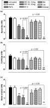
PIP-18 modulates joint inflammation and bone destruction more favorably than AF-2 peptide and DMARDs. Differential histologic scores (HS) of ankle joints of untreated Tg197 mice or those treated with the peptides (P-NT.II and phospholipase inhibitor from python (PIP)-18) or comparator drugs (methotrexate (Mtx); celecoxib (Cxb); infliximab (infxmab); antiflammin-2 (AF-2)) are shown. Compared with other drugs, infliximab and the peptides P-NT.II and PIP-18 significantly inhibited (a) synovitis, (b) cartilage destruction and (c) bone erosion. DMARD = disease-modifying anti-rheumatic drug.
Figure 8
Figure 8
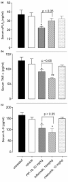
Serum levels of murine sPLA2 and IL-6, and human TNF-α. Tg197 mice received either vehicle (0.5% dimethyl sulfoxide in phosphate-buffered saline), peptides (P-NT.II or PIP-18), or comparator drugs (antiflammin-2, methotrexate, celecoxib and infliximab) at age three weeks (disease onset), and blood samples collected by cardiac puncture at termination (age eight weeks). Murine (m) serum secretory phospholipase A2 (sPLA2) levels were measured with an Escherichia coli membrane assay. Analysis of murine TNF-α and IL-6 was done by ELISA. Values are the mean ± standard error of the mean of each group; *P < 0.05; **P < 0.01 vs untreated or vehicle treated Tg197 mice.
Figure 9
Figure 9
Possible mechanism of PIP-18 suppression on IL-stimulated expression of sPLA2 and MMPs. IL-1β and/or TNF initiate the expression of secretory phospholipase A2 (sPLA2)-IIA and matrix metalloproteinases (MMP) through activation of mitogen-activated protein kinase (MAPK) cascade. (1) phospholipase inhibitor from python (PIP)-18 blocks p38 MAPK phosphorylation and reduces activation of transcription factors (activator protein-1 (AP-1), activating transcription factor 2 (ATF-2)), which regulate the transcription of sPLA2-IIA, MMPs (MMP-1, MMP-2, MMP-3, MMP-9) and proinflammatory cytokines (IL-6, TNF, IL-1). This results in downregulation of these genes and decreased protein secretions. (2) Inhibition of sPLA2 enzymatic activity by PIP-18 contributes to reduced generation of arachidonic acid for prostaglandin production. MAPKKK = MAPK kinase kinase; MAPKK = MAPK kinase; PGE2 = prostaglandin E2; sPLA2-IIA = secretory phospholipase A2-Group IIA; solid arrows, known pathways; ┤, inhibition (NF-κB pathway is not shown here).

Similar articles

Cited by

References

    1. Bongartz TA, Sutton J, Sweeting MJ, Buchan I, Matteson EL, Montori V. Anti-TNF antibody therapy in rheumatoid arthritis and the risk of serious infections and malignancies: systematic review and meta-analysis of rare harmful effects in randomized controlled trials. JAMA. 2006;295:2275–2285. doi: 10.1001/jama.295.19.2275. - DOI - PubMed
    1. Smolen JS, Aletaha D, Koeller M, Weisman MH, Emery P. New therapies for treatment of rheumatoid arthritis. Lancet. 2007;370:1861–1874. doi: 10.1016/S0140-6736(07)60784-3. - DOI - PubMed
    1. Mohammed FF, Smookler DS, Khokha R. Metalloproteinases, inflammation, and rheumatoid arthritis. Ann Rheum Dis. 2003;62 Suppl 2:ii43–ii47. - PMC - PubMed
    1. Masuda S, Murakami M, Komiyama K, Ishihara M, Ishikawa Y, Ishii T, Kudo I. Various secretory phospholipase A2 enzymes are expressed in rheumatoid arthritis and augment prostaglandin production in cultured synovial cells. FEBS J. 2005;272:655–672. doi: 10.1111/j.1742-4658.2004.04489.x. - DOI - PubMed
    1. Yedgar S, Cohen Y, Shoseyov D. Control of phospholipase A2 activities for the treatment of inflammatory conditions. Biochim Biophys Acta. 2006;1761:1373–1382. - PubMed

Publication types

MeSH terms