. 2009 Dec;30(35):6748-56.
doi: 10.1016/j.biomaterials.2009.08.042. Epub 2009 Sep 17.

Magnetic resonance imaging of multifunctional pluronic stabilized iron-oxide nanoparticles in tumor-bearing mice

Affiliations

Magnetic resonance imaging of multifunctional pluronic stabilized iron-oxide nanoparticles in tumor-bearing mice

Tapan K Jain et al. Biomaterials. 2009 Dec.

Abstract

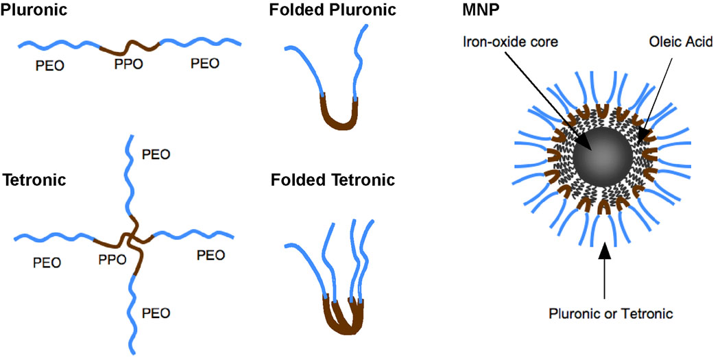
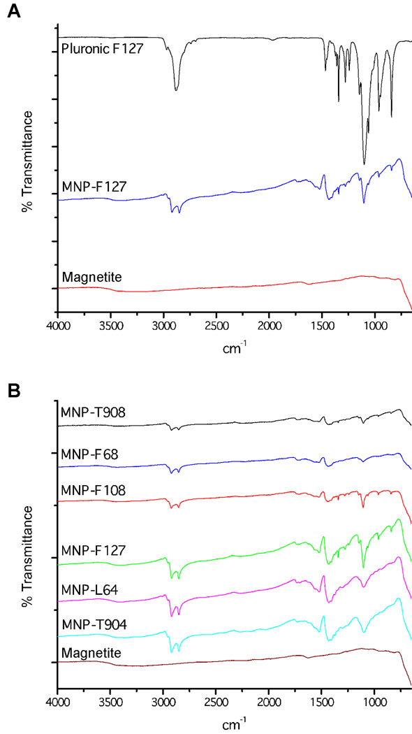
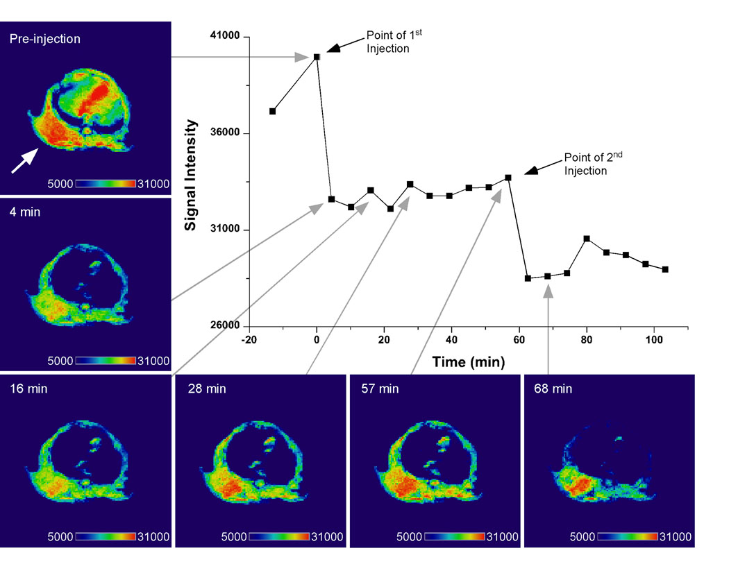
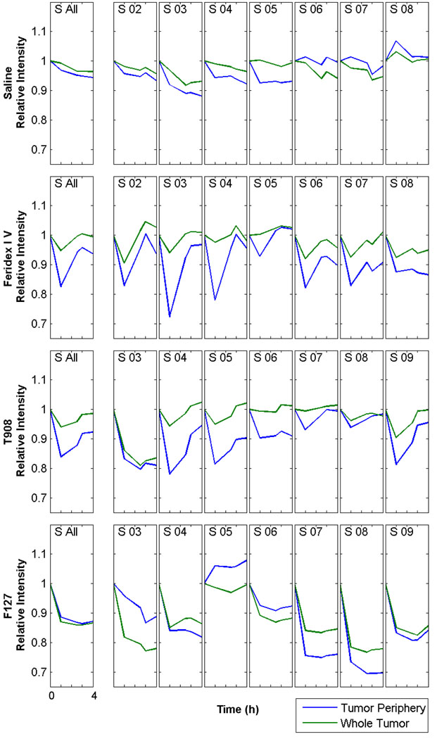
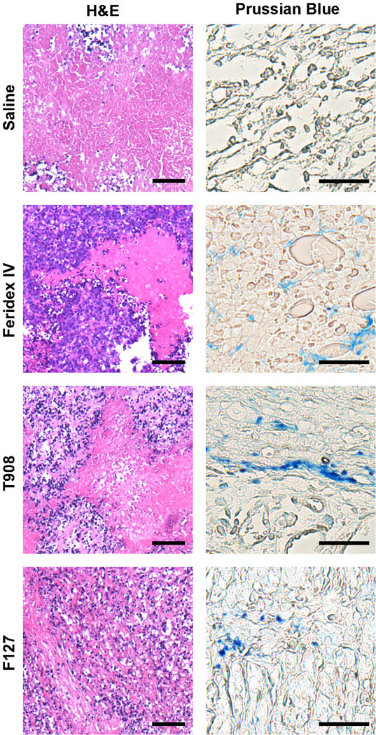
We are investigating the magnetic resonance imaging characteristics of magnetic nanoparticles (MNPs) that consist of an iron-oxide magnetic core coated with oleic acid (OA), then stabilized with a pluronic or tetronic block copolymer. Since pluronics and tetronics vary structurally, and also in the ratio of hydrophobic (poly[propylene oxide]) and hydrophilic (poly[ethylene oxide]) segments in the polymer chain and in molecular weight, it was hypothesized that their anchoring to the OA coating around the magnetic core could significantly influence the physical properties of MNPs, their interactions with biological environment following intravenous administration, and ability to localize to tumors. The amount of block copolymer associated with MNPs was seen to depend upon their molecular structures and influence the characteristics of MNPs. Pluronic F127-modified MNPs demonstrated sustained and enhanced contrast in the whole tumor, whereas that of Feridex IV was transient and confined to the tumor periphery. In conclusion, our pluronic F127-coated MNPs, which can also be loaded with anticancer agents for drug delivery, can be developed as an effective cancer theranostic agent, i.e. an agent with combined drug delivery and imaging properties.


Figures

Fig. 1
Fig. 1
Schematic of MNPs. Each particle contains an iron-oxide core coated with OA and is coated with either pluronic (single PEO-PPO-PEO subunit) or tetronic (two PEO-PPO-PEO subunits). The PPO subunit from the copolymers adsorbs onto the OA rendering the MNPs dispersible in aqueous solution. (Pluronic and tetronic are registered trademarks of BASF SE, Ludwigshafen, Germany.)
Fig. 2
Fig. 2
Characterization of anchoring of pluronic or tetronic to the OA coated MNP. A, FTIR peaks characteristic for pluronic F127 are present on F127-modified MNPs and not OA-magnetite core confirming adsorption of the pluronic to the magnetite core. B, Peaks for the C-O-C and CH2 functional groups of pluronic and tetronic copolymers confirm that each of the six copolymers anchors onto the magnetite particle.
Fig. 3
Fig. 3
T2-weighted image of tumor bearing mouse injected with pluronic F127-modifed MNPs. Enhanced contrast in the tumor (denoted by arrow) is apparent 4 min after the initial injection and is more pronounced at 68 min after a second injection of the MNPs. Images were analyzed for signal intensity in the tumor with Amira software (Visage Imaging, Inc., San Diego, CA).
Fig. 4
Fig. 4
MNPs taken up within the tumor tissue enhance MRI contrast. Contrast enhancement within the whole tumor and vascular tumor periphery for mice injected with saline, Feridex IV, F127-modified MNPs, or T908-modifed MNPs. A single ROI was drawn around the tumor at each axial slice (S 02–S 09) for the pre-injection image (0 h) and the signal intensity quantified. The same ROI was used to calculate the signal intensity at 1, 2.5, 3 and 4 h. The signal intensity from a second ROI drawn just inside the first was subtracted from the first to determine the signal intensity within the tumor periphery. An area weighted average of the slices gave the signal intensity for the whole tumor (S All). X-axis: each tick mark represents 1 h, beginning at 0 h and ending 4 h post injection of saline or the iron-oxide contrast agent. Images analyzed in MATLAB (The MathWorks, Natick, MA). Data represented from at least two repeats.
Fig. 5
Fig. 5
Tumor histological analysis after injection with iron-oxide contrast agents. Blue-violet staining of the viable periphery and red-pink staining of the necrotic core is evident in each H&E-stained tumor section. Iron from the MNPs stained blue in the tumor periphery 24 h after MNP injection. The scale bars are 25 µm in all H&E-stained images, and 10 µm in all Prussian Blue-stained images.

References

    1. Sosnovik DE, Nahrendorf M, Weissleder R. Magnetic nanoparticles for MR imaging: agents, techniques and cardiovascular applications. Basic Res Cardiol. 2008;103:122–130. - PMC - PubMed
    1. Weber MA, Giesel FL, Stieltjes B. MRI for identification of progression in brain tumors: from morphology to function. Expert Rev Neurother. 2008;8:1507–1525. - PubMed
    1. Khoo VS, Joon DL. New developments in MRI for target volume delineation in radiotherapy. Br J Radiol. 2006;79(Spec No 1):S2–S15. - PubMed
    1. Frangioni JV. New Technologies for Human Cancer Imaging. J Clin Oncol. 2008;26:4012–4021. - PMC - PubMed
    1. Xie J, Xu C, Kohler N, Hou Y, Sun S. Controlled PEGylation of monodisperse Fe3O4 nanoparticles for reduced non-specific uptake by macrophage cells. Adv Mater. 2007;19:3163–3166.

Publication types