. 2009 Sep 29;19(18):1511-8.
doi: 10.1016/j.cub.2009.07.069. Epub 2009 Sep 17.

Cell-cycle control by physiological matrix elasticity and in vivo tissue stiffening

Affiliations

Cell-cycle control by physiological matrix elasticity and in vivo tissue stiffening

Eric A Klein et al. Curr Biol. .

Abstract

Background: A number of adhesion-mediated signaling pathways and cell-cycle events have been identified that regulate cell proliferation, yet studies to date have been unable to determine which of these pathways control mitogenesis in response to physiologically relevant changes in tissue elasticity. In this report, we use hydrogel-based substrata matched to biological tissue stiffness to investigate the effects of matrix elasticity on the cell cycle.

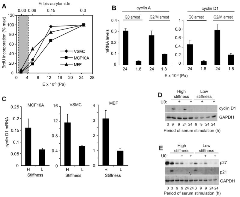
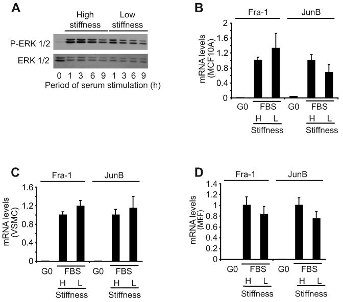
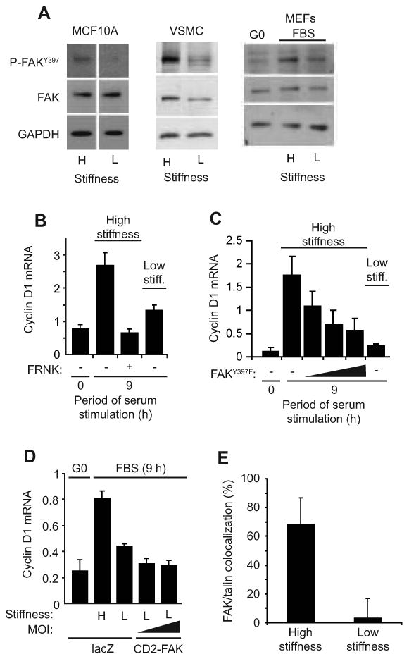
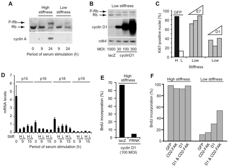
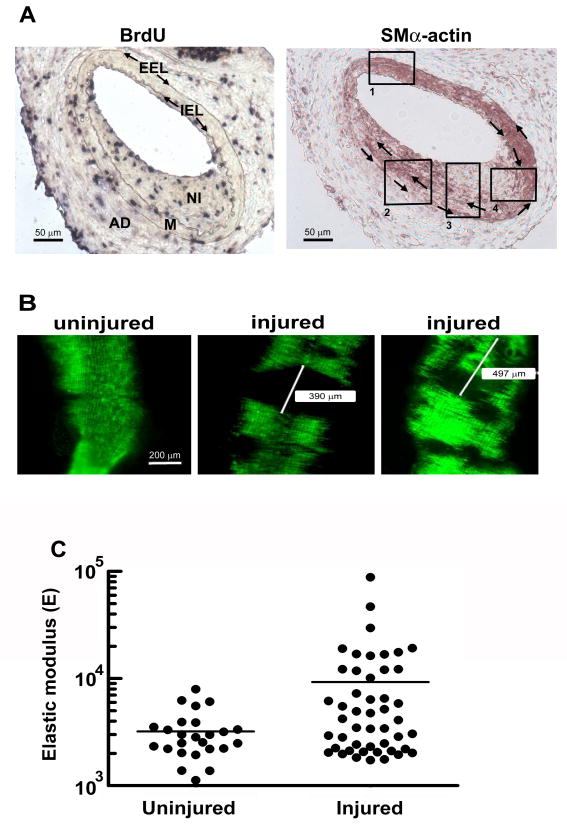
Results: We find that physiological tissue stiffness acts as a cell-cycle inhibitor in mammary epithelial cells and vascular smooth muscle cells; subcellular analysis in these cells, mouse embryonic fibroblasts, and osteoblasts shows that cell-cycle control by matrix stiffness is widely conserved. Remarkably, most mitogenic events previously documented as extracellular matrix (ECM)/integrin-dependent proceed normally when matrix stiffness is altered in the range that controls mitogenesis. These include ERK activity, immediate-early gene expression, and cdk inhibitor expression. In contrast, FAK-dependent Rac activation, Rac-dependent cyclin D1 gene induction, and cyclin D1-dependent Rb phosphorylation are strongly inhibited at physiological tissue stiffness and rescued when the matrix is stiffened in vitro. Importantly, the combined use of atomic force microscopy and fluorescence imaging in mice shows that comparable increases in tissue stiffness occur at sites of cell proliferation in vivo.

Conclusions: Matrix remodeling associated with pathogenesis is in itself a positive regulator of the cell cycle through a highly selective effect on integrin-dependent signaling to FAK, Rac, and cyclin D1.


Figures

Figure 1
Figure 1. Selective effect of ECM stiffness on cyclin D1 gene expression
(A) Serum-starved cells were stimulated with mitogens, incubated with BrdU, and reseeded on hydrogels made with a constant 7.5% acrylamide. Bis-acrylamide varied from 0.03%-0.3%. After 24 h (MCF10A cells and MEFs) or 48 h (VSMCs), the cells were fixed and BrdU incorporation was determined. The graph compiles results from an individual experiment for each cell type and shows percent maximal BrdU incorporation compared to the stiffest hydrogel. The shaded area highlights the range of elastic moduli measured in mouse mammary glands and arteries as determined by milliprobe indentation and AFM; see Suppl. Table 2. (B) MEFs were synchronized at G0 (by 48 h serum-starvation) or G2/M (by treatment with 5 μg/ml nocodozole for 24 h). The cells were reseeded on hydrogels and stimulated with 10% FBS. RNA was isolated 24 h after reseeding and analyzed by QPCR for cyclin A or cyclin D1 mRNAs. (C) Serum-starved cells were reseeded on low (L) and high (H) stiffness hydrogels with mitogens. Cyclin D1 mRNA was measured by QPCR at times corresponding to optimal induction (12 h for MCF10A cells, 24 h for VSMCs, and 9 h for MEFs). (D and E) Serum-starved MEFs were pre-treated with DMSO (vehicle) or U0126 (U0) prior to reseeding on hydrogels and stimulation with 10% FBS. Reseeded cells were collected at the indicated times and analyzed by western blotting.
Figure 2
Figure 2. Regulation of mitogenesis and cyclin D1 gene expression by ECM stiffness is ERK-independent
(A) Serum-starved MEFs were reseeded on hydrogels and stimulated with 10% FBS. Reseeded cells were collected at the indicated times and analyzed by western blotting. (B-D) Serum-starved cells were reseeded on high (H) and low (L) stiffness hydrogels with mitogens. RNA was collected from quiescent cells (G0) and cells stimulated with 10% FBS at times optimal for JunB and Fra1 mRNA induction (1 and 3 h, respectively) in the three cell types. Fra-1 and JunB mRNA levels were determined by QPCR, normalized to 18S rRNA, and plotted relative to the levels of the high stiffness samples.
Figure 3
Figure 3. FAK is linked to stiffness-dependent induction of cyclin D1 mRNA
(A) Serum-starved cells were reseeded on high (H) and low (L) stiffness hydrogels with mitogens, collected 3 hr after reseeding, and analyzed by western blotting. The white vertical line in the MCF10A blot indicates where extraneous information was removed from the blot. (B) Starved MEFs infected with adenoviruses encoding LacZ (control; -) or GFP-FRNK were reseeded on hydrogels with 10% FBS. Cyclin D1 mRNA was measured by QPCR. (C) The experiment in panel B was repeated using an adenovirus encoding FAKY397F (at 250, 500, and 1500 MOI) rather than FRNK. (D) Starved MEFs infected with adenoviruses encoding LacZ or CD2-FAK (50 and 100 MOI) were incubated on hydrogels with 10% FBS, and cyclin D1 mRNA was measured by QPCR. CD2-FAK percent infection was 70-90% as determined from its GFP IRES. (E) Asynchronous MEFs were reseeded on hydrogels. Cells were fixed 24 h after reseeding and immunostained for FAK and talin. FAK and talin colocalization was determined as described in Experimental Procedures and the legend to Suppl. Fig. 9. Results are plotted as mean colocalization ± s.d, p<10-6.
Figure 4
Figure 4. Matrix stiffness regulates the FAK-Rac-cyclin D1 signaling pathway
(A-D) Serum-starved MEFs were infected with adenoviruses encoding GFP (control), RacN17, RacV12, FRNK, or FAKY397F. Cells were reseeded on high (H) and low (L) stiffness hydrogels and stimulated with 10% FBS for 9 h to measure induction of cyclin D1 mRNA by QPCR (panels A and D) or 30 min to measure Rac GTP-loading by G-LISA (panels B and C). The data in panel D show the mean and SEM of 3 experiments. Statistical significance was determined by t-test (* p=0.029, ** p=0.015, # not significant p=0.19). (E) The experiment in panel D was repeated except that the cells were incubated with BrdU and fixed at 24 h for the determination of S phase entry.
Figure 5
Figure 5. Matrix stiffness regulates cyclin D1 function downstream of its expression
(A) Starved MEFs were reseeded on hydrogels with 10% FBS. Cell lysates were collected and analyzed by western blotting. (B) Starved MEFs infected with adenoviruses encoding LacZ or cyclin D1 (30-300 MOI) were reseeded on hydrogels with 10% FBS. Cells were collected at 24 h and analyzed by western blotting. (C) Starved MEFs infected with adenoviruses encoding GFP (1000 MOI), cyclin D1 (100, 300, and 1000 MOI), or HPV-E7 (100, 300, and 1000 MOI) were reseeded on high (H) and low (L) stiffness hydrogels and incubated with 10% FBS. S phase entry was measured 24 h after plating by immunostaining for Ki-67. (D) Starved MEFs were reseeded on hydrogels with 10% FBS. INK4 mRNA levels were measured by QPCR. (E) Starved MEFs infected with adenoviruses encoding cyclin D1 (100 MOI) and either LacZ or GFP-FRNK were reseeded in 10% FBS with BrdU. Cells were fixed at 24 h for analysis of BrdU incorporation. (F) MEFs infected with adenoviruses encoding GFP, CD2-FAK, cyclin D1 (100 MOI), or cyclin D1 & CD2-FAK were starved and plated on the high and low stiffness substrata and stimulated with 10% FBS for 24 h; S phase entry was determined by BrdU incorporation.
Figure 6
Figure 6. In vivo arterial stiffening at sites of cell proliferation
Male, 6-mo C57BL/6 mice were subjected to fine-wire femoral artery injury and given BrdU. (A) The external and internal elastic laminae (EEL and IEL, respectively) were visualized by elastin staining, and overall arterial morphology was determined by staining with hematoxylin-eosin. These procedures allowed for the identification of the adventitia (AD), the medial layer of VSMCS (M) and the site of injury (neointima; NI). Adjacent sections were analyzed by immunohistochemistry with anti-BrdU (Roche, 1299964) to visualize proliferating cells (left) and anti-α-smooth muscle actin (clone 1A4, Sigma) to visualize regions of differentiated and de-differentiated VSMCs (right). Clockwise and counterclockwise arrows in the right panel mark the IEL and EEL, respectively. Boxed regions highlight distinct patterns of VSMC differentiation in the media and neointima. (B) Male, 6-mo SMA-GFP mice were subjected to fine-wire femoral artery injury. Uninjured and contralateral injured arteries were isolated, carefully opened, and imaged for GFP-fluorescence. Representative areas of the uninjured and injured arteries are shown for a single mouse. Scale bar = 200 μm. (C) Uninjured (control) and injured femoral arteries from SMA-GFP mice were collected 2 weeks after the fine-wire injury procedure. AFM was used to measure the elastic modulus of several GFP positive regions of uninjured arteries and GFP-negative regions of injured arteries. The figure compiles AFM measurements obtained from 4 mice. p=0.0029 (one-way t-test with Welch correction for unequal variance; p for unequal variance <0.0001).

References

    1. Assoian RK, Schwartz MA. Coordinate signaling by integrins and receptor tyrosine kinases in the regulation of G1 phase cell-cycle progression. Curr Opin Genet Dev. 2001;11:48–53. - PubMed
    1. Fringer J, Grinnell F. Fibroblast quiescence in floating or released collagen matrices: contribution of the ERK signaling pathway and actin cytoskeletal organization. J Biol Chem. 2001;276:31047–31052. - PubMed
    1. Rosenfeldt H, Grinnell F. Fibroblast quiescence and the disruption of ERK signaling in mechanically unloaded collagen matrices. J Biol Chem. 2000;275:3088–3092. - PubMed
    1. Koyama H, Raines EW, Bornfeldt KE, Roberts JM, Ross R. Fibrillar collagen inhibits arterial smooth muscle proliferation through regulation of Cdk2 inhibitors. Cell. 1996;87:1069–1078. - PubMed
    1. Wall SJ, Zhong ZD, DeClerck YA. The cyclin-dependent kinase inhibitors p15INK4B and p21CIP1 are critical regulators of fibrillar collagen-induced tumor cell cycle arrest. J Biol Chem. 2007;282:24471–24476. - PubMed

Publication types