. 2009 Nov 27;389(4):657-62.
doi: 10.1016/j.bbrc.2009.09.049. Epub 2009 Sep 17.

Cloning and characterization of the 5'UTR of the rat anti-apoptotic Bcl-w gene

Affiliations

Cloning and characterization of the 5'UTR of the rat anti-apoptotic Bcl-w gene

Martine Uittenbogaard et al. Biochem Biophys Res Commun. .

Abstract

The anti-apoptotic Bcl-w regulator, which is expressed in the developing and mature brain, not only promotes neuronal survival, but also neuronal differentiation. However, its transcriptional regulation remains to be elucidated due to a lack of knowledge of the Bcl-w promoter. Here, we report the mapping and characterization of the rat Bcl-w promoter, which is highly conserved between the human, mouse, and rat species. Using a series of 5' and 3' deletions, we mapped the TATA-less minimal Bcl-w promoter and showed that it is under a combinatorial regulation with the neurogenic bHLH transcription factor NeuroD6 mediating its activation, validating our previous finding of increased expression of the Bcl-w protein in stably transfected PC12-NeuroD6 cells. Upon stress, NeuroD6 promotes colocalization of Bcl-w with mitochondria and endoplasmic reticulum. Finally, we provide the first evidence of Bcl-w localization in the growth cones of differentiating neuronal cells, suggestive of a potential synaptic neuroprotective role.


Figures

Fig. 1
Fig. 1
Constitutive expression of NeuroD6 induces expression of the Bcl-w protein at level similar to that of NGF-induced neuronal differentiation of PC12 cells. Cell lysates from untreated PC12-ND6 cells and NGF-treated PC12 cells were analyzed by Western blot using an anti-Bcl-w polyclonal antibody. Equal loading was verified using an anti-GAPDH antibody.
Fig. 2
Fig. 2
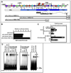
The rat Bcl-w promoter is TATA-less and contains multiple transcription start sites. (A). Primer extension of the rat Bcl-w promoter during neuronal differentiation. Poly (A) RNA isolated from adult rat brain, untreated and NGF-treated PC12 cells was annealed to an end-labeled primer. The arrows indicate major transcription start sites found in common with adult rat brain and NGF-PC12 cells, while the star symbol indicates minor transcription start sites specific to adult rat brain. (B). Mapping of the multiple Bcl-w transcription start sites and exon boundaries by 5’RACE. We identified a novel alternative non-coding exon 1b in the 5’UTR of the rat Bcl-w gene. Transcription start sites mapping in the non-coding exons 1a and 1b are indicated with blue and red arrows, respectively. Donor splice sites (SD) of non-coding exons 1a and 1b are indicated by a blue and red vertical line, respectively.
Fig. 3
Fig. 3
Regulation of the rat Bcl-w promoter. (A). Map of the rat Bcl-w promoter and Bcl-w luciferase reporter constructs. The schematic representation illustrates the degree of homology between human, mouse, and rat genomic sequences. The numbering is relative to the translation start site (+1). Multiple transcription start sites span from −750 to −870, with clustered Sp1 binding sites indicated by red arrows. Relevant conserved enhancer elements are schematically represented by different symbols with their names indicated below. Asterisks indicate enhancer elements conserved in the three species, while the arrow indicates the protected E3 E-box as shown by EMSA. (B). Bcl-w promoter activity in PC12 cells treated with NGF (50 ng/ml) for three days. Results are expressed as means ± SD from 4 independent experiments done in triplicate. (C). NeuroD6 upregulates the Bcl-w promoter activity. PC12 and PC12-ND6 cells were transfected with the Bcl-w (−1800/+1) reporter construct, with or without eukaryotic expression vector containing NeuroD6, NeuroD6-mut1, or Id2. Cells were either untreated or treated with NGF for 3 days. Results are expressed as means ± SD of fold of induction relative to the Bcl-w (−1800/+1) reporter construct from 4 independent experiments done in triplicate. (D). The E3 E-box is occupied by nuclear proteins from both untreated PC12-ND6 cells and NGF-PC12 cells. Nuclear extracts (8 µg) were incubated in the presence of increasing amounts specific (left panel), mutated (middle panel) competitors or pre-immune IgG or purified anti-NeuroD6 IgG (right panel).
Fig. 4
Fig. 4
Subcellular localization of Bcl-w protein in the absence or presence of stress stimulus. (A). Immunocytochemistry was performed on serum-grown (t=0) and serum-deprived (t=48 hr) PC12-ND6 cells to examine Bcl-w subcellular localization (green) and its overlapping expression with mitochondria and endoplasmic reticulum (ER), using COXV α subunit as a mitochondrial marker (red) and poly-D-isomerase (PDI) as an ER marker (red). Cells were incubated with the nuclear counterstain DAPI (blue). Scale bars are indicated at the bottom right corner of merged images. Magnification of the soma and growth cone is shown on right panels. Colocalization between Bcl-w and mitochondria and ER is indicated by arrows, while Bcl-w expression in the vicinity of either organelle is indicated by arrowheads. (B). Expression levels of Bcl-w protein were analyzed by immunoblot analysis using mitochondria-enriched fractions isolated from serum grown (t= 0) or serum-deprived (t=2d) PC12-ND6 cells. Equal loading was verified using an antibody against subunit α of COX V.
Fig. 4
Fig. 4
Subcellular localization of Bcl-w protein in the absence or presence of stress stimulus. (A). Immunocytochemistry was performed on serum-grown (t=0) and serum-deprived (t=48 hr) PC12-ND6 cells to examine Bcl-w subcellular localization (green) and its overlapping expression with mitochondria and endoplasmic reticulum (ER), using COXV α subunit as a mitochondrial marker (red) and poly-D-isomerase (PDI) as an ER marker (red). Cells were incubated with the nuclear counterstain DAPI (blue). Scale bars are indicated at the bottom right corner of merged images. Magnification of the soma and growth cone is shown on right panels. Colocalization between Bcl-w and mitochondria and ER is indicated by arrows, while Bcl-w expression in the vicinity of either organelle is indicated by arrowheads. (B). Expression levels of Bcl-w protein were analyzed by immunoblot analysis using mitochondria-enriched fractions isolated from serum grown (t= 0) or serum-deprived (t=2d) PC12-ND6 cells. Equal loading was verified using an antibody against subunit α of COX V.

References

    1. Oppenheim RW. Cell death during development of the nervous system. Annu. Rev. 1991;14:453–501. - PubMed
    1. Putcha GV, Johnson EM., Jr Men are but worms: neuronal cell death in C. elegans and vertebrates. Cell Death Differ. 2004;11:38–48. - PubMed
    1. Benn SC, Woolf CJ. Adult neuron survival strategies- slamming on the brakes. Nature Rev. Neurosci. 2004;5:686–700. - PubMed
    1. Liu M, Pereira FA, Price SD, Chu M-J, Shope C, Himes D, Eatock RA, Brownell WE, Lysakowski A, Tsai MJ. Essential role of Beta2/NeuroD1 in development of the vestibular and auditory systems. Genes & Dev. 2000;14:2839–2854. - PMC - PubMed
    1. Schwab MH, Bartholomae A, Heimrich B, Feldmeyer D, Druffel-Augustin S, Goebbels S, Naya FJ, Zhao S, Frotscher M, Tsai MJ, Nave KA. Neuronal basic helix-loop-helix proteins (NEX and BETA2/Neuro D) regulate terminal granule cell differentiation in the hippocampus. J Neurosci. 2000;20:3714–3724. - PMC - PubMed

Publication types