

T cell extravasation: demonstration of synergy between activation of CXCR3 and the T cell receptor
- PMID: 19767105
- PMCID: PMC2817451
- DOI: 10.1016/j.molimm.2009.08.021
T cell extravasation: demonstration of synergy between activation of CXCR3 and the T cell receptor
Abstract
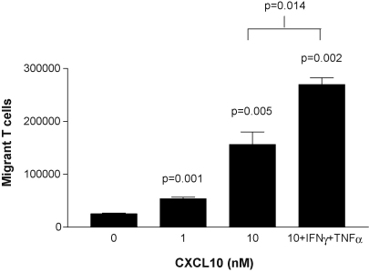
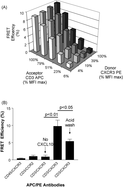
Endothelial cells present chemokines to T cells and can also stimulate the T cell antigen receptor by presentation of peptide-MHC antigen complexes. This study was designed to investigate the potential synergy between stimulation of the chemokine receptor CXCR3 and the human T cell receptor complex. Transendothelial T cell migration towards CXCL10 was modified by crosslinking CD3 immediately before addition to the endothelium. When resting endothelium was used, T cells which had been activated by crosslinking CD3 for only 1 min showed a significant reduction (p<0.0001) in migration when compared with untreated T cells. By contrast, endothelial cells which had been activated by stimulation with interferon-gamma and tumour necrosis factor-alpha supported a specific increase in the migration of activated T cells; this was most apparent after CD3 had been activated for 90 min (p<0.0001). The molecular basis for synergy between CXCR3 and the T cell receptor complex was investigated by measurement of fluorescence resonance energy transfer. This showed that CXCL10 induced a close (<10 nm) spatial association between CXCR3 and the CD3epsilon subunit on the cell-surface. These data demonstrate that stimulation of both CXCR3 and the T cell receptor has the potential to enhance specifically both the proliferation and extravasation of specific T cells during episodes of local inflammation.
Figures

Similar articles
-
CXCR3 internalization following T cell-endothelial cell contact: preferential role of IFN-inducible T cell alpha chemoattractant (CXCL11).J Immunol. 2001 Dec 15;167(12):7084-93. doi: 10.4049/jimmunol.167.12.7084. J Immunol. 2001. PMID: 11739530
-
Human mesenchymal stem cells target adhesion molecules and receptors involved in T cell extravasation.Stem Cell Res Ther. 2015 Dec 10;6:245. doi: 10.1186/s13287-015-0222-y. Stem Cell Res Ther. 2015. PMID: 26651832 Free PMC article.
-
The relative activity of CXCR3 and CCR5 ligands in T lymphocyte migration: concordant and disparate activities in vitro and in vivo.J Leukoc Biol. 2003 Nov;74(5):791-9. doi: 10.1189/jlb.1102547. Epub 2003 Aug 1. J Leukoc Biol. 2003. PMID: 12960247
-
CXCL9, CXCL10, CXCL11/CXCR3 axis for immune activation - A target for novel cancer therapy.Cancer Treat Rev. 2018 Feb;63:40-47. doi: 10.1016/j.ctrv.2017.11.007. Epub 2017 Nov 26. Cancer Treat Rev. 2018. PMID: 29207310 Free PMC article. Review.
-
Chemokine (C-X-C motif) ligand (CXCL)10 in autoimmune diseases.Autoimmun Rev. 2014 Mar;13(3):272-80. doi: 10.1016/j.autrev.2013.10.010. Epub 2013 Nov 2. Autoimmun Rev. 2014. PMID: 24189283 Review.
Cited by
-
Self-assembled and perfusable microvasculature-on-chip for modeling leukocyte trafficking.Lab Chip. 2024 Jan 17;24(2):292-304. doi: 10.1039/d3lc00719g. Lab Chip. 2024. PMID: 38086670 Free PMC article.
-
Concomitant active tuberculosis prolongs survival in non-small cell lung cancer: a study in a tuberculosis-endemic country.PLoS One. 2012;7(3):e33226. doi: 10.1371/journal.pone.0033226. Epub 2012 Mar 16. PLoS One. 2012. PMID: 22438899 Free PMC article.
-
Inflammatory chemokine receptors regulate CD8(+) T cell contraction and memory generation following infection.J Exp Med. 2011 Aug 1;208(8):1621-34. doi: 10.1084/jem.20102110. Epub 2011 Jul 25. J Exp Med. 2011. PMID: 21788409 Free PMC article.
-
High IP-10 levels decrease T cell function in HIV-1-infected individuals on ART.J Leukoc Biol. 2014 Dec;96(6):1055-63. doi: 10.1189/jlb.3A0414-232RR. Epub 2014 Aug 25. J Leukoc Biol. 2014. PMID: 25157027 Free PMC article.
-
Overview of the Mechanisms that May Contribute to the Non-Redundant Activities of Interferon-Inducible CXC Chemokine Receptor 3 Ligands.Front Immunol. 2018 Jan 15;8:1970. doi: 10.3389/fimmu.2017.01970. eCollection 2017. Front Immunol. 2018. PMID: 29379506 Free PMC article. Review.
References
-
- Adams D.H., Shaw S. Leucocyte-endothelial interactions and regulation of leucocyte migration. Lancet. 1994;343:831–836. - PubMed
-
- Bacon K.B., Premack B.A., Gardner P., Schall T.J. Activation of dual T cell signaling pathways by the chemokine RANTES. Science. 1995;269:1727–1730. - PubMed
-
- Batard P., Szollosi J., Luescher I., Cerottini J.C., MacDonald R., Romero P. Use of phycoerythrin and allophycocyanin for fluorescence resonance energy transfer analyzed by flow cytometry: advantages and limitations. Cytometry. 2002;48:97–105. - PubMed
Publication types
MeSH terms
Substances
Grants and funding
LinkOut - more resources
Full Text Sources
Research Materials
