. 2010 Aug;43(2):161-72.
doi: 10.1165/rcmb.2009-0031OC. Epub 2009 Sep 18.

Endothelial-mesenchymal transition in bleomycin-induced pulmonary fibrosis

Affiliations

Endothelial-mesenchymal transition in bleomycin-induced pulmonary fibrosis

Naozumi Hashimoto et al. Am J Respir Cell Mol Biol. 2010 Aug.

Abstract

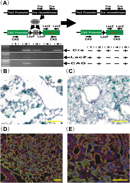
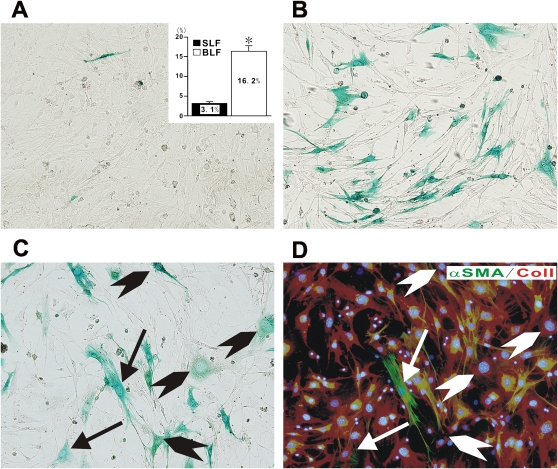
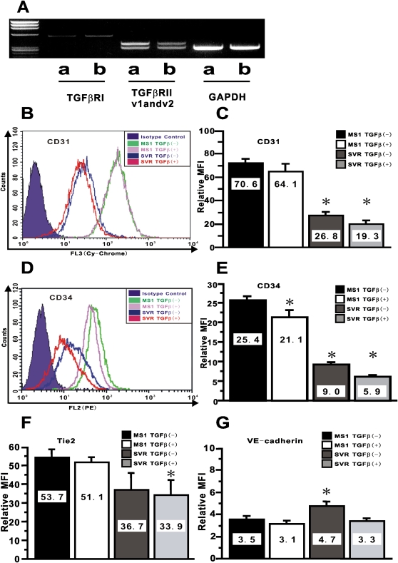
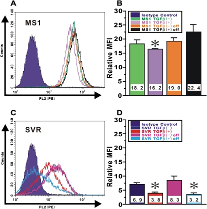
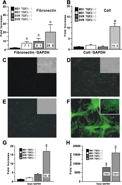
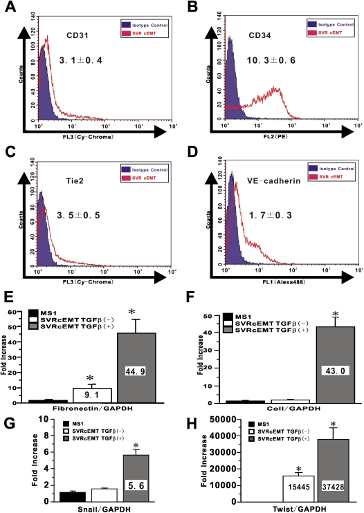
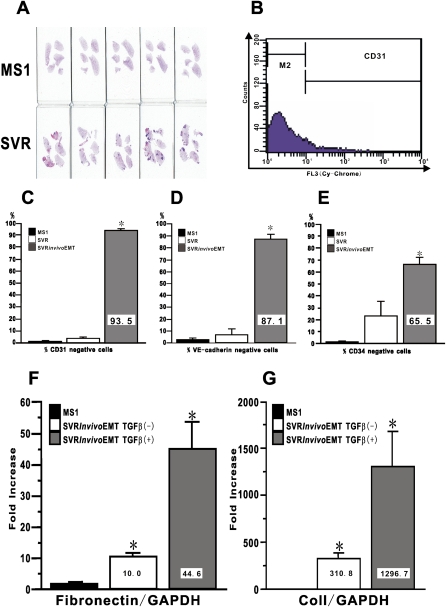
The pathological hallmark lesions in idiopathic pulmonary fibrosis are the fibroblastic foci, in which fibroblasts are thought to be involved in the tissue remodeling, matrix deposition, and cross-talk with alveolar epithelium. Recent evidence indicates that some fibroblasts in fibrosis may be derived from bone marrow progenitors as well as from epithelial cells through epithelial-mesenchymal transition. To evaluate whether endothelial cells could represent an additional source for fibroblasts, bleomycin-induced lung fibrosis was established in Tie2-Cre/CAG-CAT-LacZ double-transgenic mice, in which LacZ was stably expressed in pan-endothelial cells. Combined X-gal staining and immunocytochemical staining for type I collagen and alpha-smooth muscle actin revealed the presence of X-gal-positive cells in lung fibroblast cultures from bleomycin-treated mice. To explore the underlying mechanisms, by which loss of endothelial-specific markers and gain of mesenchymal phenotypes could be involved in microvascular endothelial cells, the effects of activated Ras and TGF-beta on the microvascular endothelial cell line MS1 were analyzed. Combined treatment with activated Ras and TGF-beta caused a significant loss of endothelial-specific markers, while inducing de novo mesenchymal phenotypes. The altered expression of these markers in MS1 cells with activated Ras persisted after withdrawal of TGF-beta in vitro and in vivo. These findings are the first to show that lung capillary endothelial cells could give rise to significant numbers of fibroblasts through an endothelial-mesenchymal transition in bleomycin-induced lung fibrosis model.


Figures

Figure 1.
Figure 1.
Generation of Tie2-Cre/CAG-CAT-LacZ double-transgenic mice (CAG mice) and bleomycin (BLM)-induced lung fibrosis model. To obtain CAG mice, Tie2-Cre Tg mice were bred with CAG-CAT-LacZ Tg mice. (A) Top: Schematic description of gene constructs in CAG mice. Bottom: Genotyping analyses. Genotyping analyses were performed for off spring of CAG mice. The representative electropherogram of Cre (left top), LacZ (left middle), and CAG (left bottom) were shown for a set of five offsprings. The results were summarized (right panel). X-gal staining was performed for the lung tissues from (B) saline- or (C) BLM- treated CAG mice at Day 28 (×400 magnification). Double immunostaining for CD31 (green) and Col I (red) was performed for the lung tissues from BLM mice (D, ×200 magnification; E, ×400 magnification). Scale bars in B–D indicate 50 μm.
Figure 2.
Figure 2.
Characterization of BLM-induced lung fibroblasts derived from CAG mice. Saline-treated lung fibroblasts (SLF) or BLM-treated lung fibroblasts (BLF) derived from CAG mice were isolated and then stained with X-gal (SLF in A and BLF in B, respectively). The percentage of X-gal–positive cells among total counted cells, based on Hoechst 33342 nucleus staining, were shown in inset in A. Data shown in inset represent the means ± SEM from at least four samples (4 for SLF and 8 for BLF, respectively) in three independent experiments. Combined staining with X-gal and sequential immucytochemistry for Col I (red) and α-SMA (green) were performed for BLF derived from CAG mice (C, X-gal staining; D, immunocytochemistry for Col I and α-SMA, respectively). Arrows indicate X-gal (+)/Col I (+)/α-SMA (+) myofibroblasts. Arrowheads indicate X-gal (+)/Col I (+)/α-SMA (−) fibroblasts. A representative example of at least three independent experiments is shown. All images were photographed at ×200 magnification.
Figure 3.
Figure 3.
Altered expression of endothelial-specific markers by combined treatment with activated Ras and TGF-β. In A, the results of RT-PCR analysis for TGF-βRI, TGF-βRII variant1 (v1), variant2 (v2), and GAPDH are shown. Data shown are representative electropherograms of the indicated products using RNA samples from: MS1 (lane a) and SVR (lane b). These are representative of three independent experiments. CD31 in B and CD34 in D on endothelial MS1 and SVR treated with vehicle or TGF-β at 10 ng/ml for 24 hours were shown as overlaid histogram by FACS. The relative mean fluorescence intensity (MFI) for CD31 in C, for CD34 in E, for Tie2 in F, and for VE-cadherin in G were calculated as described in the online supplement. Data shown represent the means ± SEM from at least three independent experiments. Asterisks signify statistically significant difference (P < 0.05) in comparison with the relative MFI for each endothelial marker on MS1 with vehicle.
Figure 4.
Figure 4.
Persistence of altered endothelial phenotype in Ras-activated endothelial cells after withdrawal of TGF-β in vitro. Endothelial MS1 and SVR treated with vehicle or TGF-β at 10 ng/ml for 24 hours were cultured in complete medium after TGF-β removal for another 24 hours. CD34 expression on (A) MS1 cells and (C) SVR cells was shown as overlaid histogram by FACS. The relative MFI for CD34 in (B) MS1 cells and (D) SVR cells was calculated as described in the online supplement. Data shown represent the means ± SEM from at least three independent experiments. Asterisks signify statistically significant difference (P < 0.05) in comparison with the relative MFI for CD34 on MS1 or SVR with vehicle.
Figure 5.
Figure 5.
De novo induced expression of mesenchymal-specific markers in endothelial cells by combined treatment with activated Ras and TGF-β. Mesenchymal markers expression in MS1 and SVR treated with vehicle or TGF-β at 10 ng/ml for 24 hours were evaluated. Fibronectin mRNA in A and Col I mRNA in B were analyzed using real-time PCR. Data shown represent the means ± SEM from three independent experiments. Cultured cells in an 8-well Lab-Tek Chamber Slide were also treated with vehicle or 10 ng/ml TGF-β in medium without FCS for 24 hours for immunocytochemistry for α-SMA. (C–F) Treated cells were stained with FITC-conjugated mouse anti-α-SMA antibody. (C) MS1 with vehicle. (D) MS1 with TGF-β. (E) SVR with vehicle. (F) SVR with TGF-β. Magnification: ×200. The insets in CE shows the light microscopic appearance of treated cells treated with each condition. Inset in F shows the cells stained with isotype-matched control IgG for α-SMA. A representative example of at least three independent experiments is shown. Snail mRNA in G and Twist mRNA in H were also analyzed using real-time PCR. Data shown represent the means ± SEM from three independent experiments. Asterisks signify statistically significant difference (P < 0.05) in comparison with the quantitative value of targeted mRNA in MS1 with vehicle.
Figure 6.
Figure 6.
Persistence of endothelial-MT phenotype in Ras-activated endothelial cells after withdrawal of TGF-β. SVR treated with TGF-β were re-plated into the new culture dish and cultured with complete medium. The collected cells were called as cEMT cells. To evaluate whether cEMT cells can retain endothelial-MT phenotype or not, CD31 in A, CD34 in B, Tie-2 in C, and VE-cadherin expression in D on cEMT cells were shown as overlaid histogram by FACS. A representative example of at least three independent experiments is shown. Mesenchymal markers expressions in cEMT cells were also evaluated. Fibronectin mRNA in E, Col I mRNA in F, Snail mRNA in G, and Twist mRNA in H were analyzed using real-time PCR. Data shown represent the means ± SEM from three independent experiments. Asterisks signify statistically significant difference (P < 0.05) in comparison with the quantitative value of targeted mRNA in MS1.
Figure 7.
Figure 7.
In vivo acquisition of endothelial-MT phenotype in Ras-activated endothelial cells. A quantity of 1 × 10⁶ cells of MS1 or SVR were inoculated subcutaneously into the flank of nude mice. (A) The lungs from MS1-treated mice (upper row; n = 5) and SVR-treated mice (lower row; n = 5) were collected at Day 28 to evaluate lung metastasis. Collected primary tumors of SVR were digested and selected with neomycin. Selected ex vivo SVR cells, which we called in vivo EMT cells, were analyzed for the expression of endothelial-specific markers by FACS. (B) We defined the population of cells in M2 gate as marker-negative cells. The percentage of marker-negative cells for (C) CD31, (D) VE-cadherin, and (E) CD34 are shown. Data shown represent the means ± SEM from three independent experiments. Fibronectin mRNA in F and Col I mRNA in G were analyzed using real-time PCR. Data shown represent the means ± SEM from three independent experiments. Asterisks signify statistically significant difference (P < 0.05) in comparison with the quantitative value of targeted endothelial markers and mRNA in MS1.

Comment in

References

    1. Gross TJ, Hunninghake GW. Idiopathic pulmonary fibrosis. N Engl J Med 2001;345:517–525. - PubMed
    1. Thannickal VJ, Toews GB, White ES, Lynch JP III, Martinez FJ. Mechanisms of pulmonary fibrosis. Annu Rev Med 2004;55:395–417. - PubMed
    1. Iwano M, Plieth D, Danoff TM, Xue C, Okada H, Neilson EG. Evidence that fibroblasts derive from epithelium during tissue fibrosis. J Clin Invest 2002;110:341–350. - PMC - PubMed
    1. Hashimoto N, Jin H, Liu T, Chensue SW, Phan SH. Bone marrow-derived progenitor cells in pulmonary fibrosis. J Clin Invest 2004;113:243–252. - PMC - PubMed
    1. Willis BC, Liebler JM, Luby-Phelps K, Nicholson AG, Crandall ED, du Bois RM, Borok Z. Induction of epithelial-mesenchymal transition in alveolar epithelial cells by transforming growth factor-beta1: potential role in idiopathic pulmonary fibrosis. Am J Pathol 2005;166:1321–1332. - PMC - PubMed

Publication types