. 2009 Oct;11(10):1212-8.
doi: 10.1038/ncb1964. Epub 2009 Sep 20.

The bacterial virulence factor InlC perturbs apical cell junctions and promotes cell-to-cell spread of Listeria

Affiliations

The bacterial virulence factor InlC perturbs apical cell junctions and promotes cell-to-cell spread of Listeria

Tina Rajabian et al. Nat Cell Biol. 2009 Oct.

Abstract

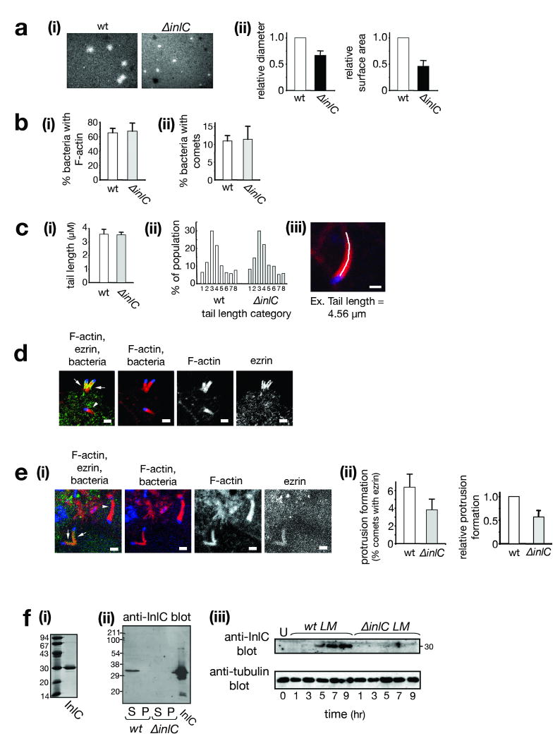
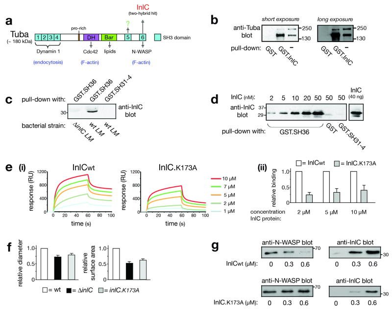
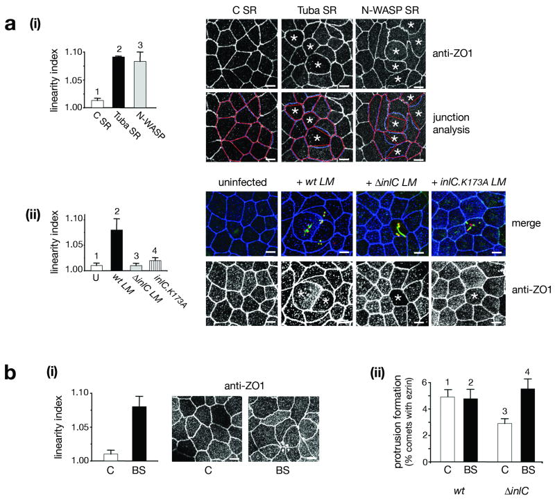
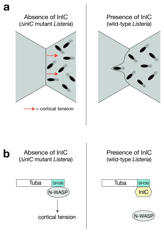
Several pathogenic bacteria, including Listeria monocytogenes, use an F-actin motility process to spread between mammalian cells. Actin 'comet tails' propel Listeria through the cytoplasm, resulting in bacteria-containing membrane protrusions that are internalized by neighbouring cells. The mechanism by which Listeria overcomes cortical tension to generate protrusions is unknown. Here, we identify bacterial and host proteins that directly regulate protrusions. We show that efficient spreading between polarized epithelial cells requires the secreted Listeria virulence protein InlC (internalin C). We next identify the mammalian adaptor protein Tuba as a ligand of InlC. InlC binds to a carboxy-terminal SH3 domain in Tuba, which normally engages the human actin regulatory protein N-WASP. InlC promotes protrusion formation by inhibiting Tuba and N-WASP activity, probably by impairing binding of N-WASP to the Tuba SH3 domain. Tuba and N-WASP are known to control the structure of apical junctions in epithelial cells. We demonstrate that, by inhibiting Tuba and N-WASP, InlC makes taut apical junctions become slack. Experiments with myosin II inhibitors indicate that InlC-mediated perturbation of apical junctions accounts for the role of this bacterial protein in protrusion formation. Collectively, our results suggest that InlC promotes bacterial dissemination by relieving cortical tension, thereby enhancing the ability of motile bacteria to deform the plasma membrane into protrusions.


Figures

Figure 1
Figure 1. InlC is needed for efficient spreading and protrusion formation in a polarized cell line
(a) InlC promotes spreading. Caco-2 BBE1 cells in transwells were infected with wild-type (wt) or ΔinlC Listeria strains. (i): Representative images of plaque assays. (ii): Average relative plaque diameters or surface areas ± s.d. (n=3). (b). InlC does not affect F-actin tail formation. (i). Average percentage (%) ± s.d. (n=3) of intracellular wild-type or ΔinlC bacteria with symmetric F-actin or comet tails. P = 0.45. (ii). Average percentage (%) ± s.d. (n=3) of intracellular bacteria with comet tails only. P = 0.46. (c) InlC does not influence tail length. (i). Average tail lengths (μm) ± s.d. (n=3). P = 0.84. (ii). Distributions of tail lengths: <1 μm (1), 1.00-1.99 μm (2), 2.00-2.99 μm (3), 3.00-3.99 μm (4), 4.00-4.99 μm (5), 5.00-5.99 μm (6), 6.00-6.99 μm (7), 7.00-14.00 μm (8). (iii). Tail length measurement. F-actin: red; bacterium: blue. Tail length is measured as a free-hand trace. Scale bar, 1 μm. (d) Ezrin localizes to protrusions. Confocal microscopy was performed on subconfluent Caco-2 BBE1 cells infected with wild-type Listeria. The left panel is a merged image showing F-actin (red), ezrin (green), and bacteria (blue). Arrows indicate ezrin-containing comet tails in protrusions projecting into space. The arrowhead indicates an ezrin-negative tail in the cell body. (e) InlC promotes protrusion formation. Scale bars, 2 μm. (i). Representative image of wild-type Listeria in cells in transwells. F-actin, ezrin, and bacteria are colored as in d. Arrows and arrowheads indicate F-actin tails containing or lacking ezrin, respectively. (ii). Quantification of ezrin recruitment. Protrusion formation is expressed as the average percentage ± s.d. (n=7) of total F-actin tails containing ezrin (left panel), or as relative values (right panel). P = 0.008. (f) InlC is expressed in infected human cells. (i) InlC used to generate antibodies. (ii) Anti-InlC Western blots of supernatants (S) or pellets (P) from broth cultures of wild-type (wt) or ΔinlC strains. (iii) Caco-2 BBE1 cells were left uninfected (U) or infected with Listeria strains for increasing times. Cell lysates were Western blotted with antibodies against InlC or tubulin.
Figure 2
Figure 2. InlC interacts with the mammalian adaptor protein Tuba
(a) Structure of Tuba. SH3, Dbl homology (DH), and Bin-Amphiphysin-Rvs (Bar) domains are indicated. The last SH3 domain (SH36) interacts with InlC or human N-WASP. Other ligands of the first four SH3 (SH31-4), DH, or Bars domain are indicated. In addition to full-length Tuba (∼180 kDa), isoforms lacking SH31-4, but retaining the DH, Bar, SH35, and SH36 domains, exist,. (b) InlC associates with ∼ 180 and ∼150 kDa Tuba isoforms from Caco-2 BBE1 cells. A GST-InlC fusion protein was incubated with cell lysates and precipitated. Precipitates were immunoblotted with antibodies against the Tuba SH36 domain. (c) The Tuba SH36 domain associates with secreted InlC. Caco-2 BBE1 cells were infected with wild-type (wt) or ΔinlC strains of L. monocytogenes (LM) for 7 h. Cell lysates were used for precipitation with GST proteins containing SH36, or the SH31-4 region as a control. (d) InlC interacts directly with SH36. GST proteins containing SH36, SH31-4, or GST alone (10 nM) were incubated with the indicated concentrations of InlC and precipitated. InlC was detected by immunoblotting. The last lane is a loading control. (e) An RxxK sequence in InlC participates in binding to SH36 (i). Binding of wild-type InlC protein (InlCwt) or InlC.K173A, as measured by SPR. Representative data from a single experiment is shown. Data from 3-4 experiments were used to estimate equilibrium dissociation constants (KD) of 9.0 ± 3.5 and 58 ± 12.3 μM for wild-type InlC and InlC.K173A, respectively. P = 0.0007. (ii). Relative binding of wild-type (wt) InlC and InlC.K173A to SH36. The data is average ± s.e.m. (n=3). (f) An RxxK sequence in InlC is needed for efficient spreading. Average relative plaque diameters (i) or surface areas (ii) ± s.d. (n=3) of wild-type (wt), ΔinlC, or inlC.K173A strains of Listeria are presented. (g) InlC displaces N-WASP from SH3-6. 10 nM GST-SH36 was incubated with 10 nM of N-WASP in the presence of the indicated concentrations of wild-type InlC (InlCwt) or InlC.K173A for 2.5 h. GST-SH36 was precipitated, and N-WASP (left panels) or InlC (right panels) detected by immunoblotting.
Figure 3
Figure 3. Tuba and N-WASP control protrusion formation
Caco-2 BBE1 cells were treated with control (C) siRNA or siRNA molecules targeting Tuba or N-WASP. About 72 h after transfection, cells were infected with wild-type (wt) or ΔinlC strains of Listeria for ∼ 5 h prior to Western blotting (a) or fixation and labeling for analysis of protrusions (b) or F-actin tails (c). (a) siRNA-mediated depletion of Tuba or N-WASP. Western blots were performed using lysates from cells treated with ‘siTuba-1’ or ‘siNWASP-1’ siRNA (Methods). Top panels display anti-Tuba or anti-N-WASP immunoblots, whereas the bottom panels show anti-tubulin blots of the stripped membranes. (b) Quantification of recruitment of ezrin to comets. Protrusion efficiency is expressed as the average percentage ± s.d. of F-actin tails that contain ezrin (Y-axis). The data are from 3-4 experiments, depending on the condition. Statistical analysis revealed significant differences (p<0.05) in the following column pairs: i+iv, iv+v, and iv+vi. *Note that the apparent difference in protrusions in cells treated with control or N-WASP siRNA (columns i and iii) is not statistically significant (p>0.05). (c) Comet tail lengths in Caco-2 BBE1 cells depleted for Tuba or N-WASP. Tail lengths (μm) are average ± s.d. values from three experiments. Statistical analysis revealed no significant difference between any of the columns (p = 0.53). The data in parts a, b, and c are from experiments using ‘siTuba-1’ or ‘siNWASP-1’ siRNA molecules (Methods). siRNA molecules that targeted different regions in Tuba or N-WASP mRNA (‘siTuba-2’ or ‘siNWASP-2; Methods) produced similar results (data not shown).
Figure 4
Figure 4. InlC, Tuba, and N-WASP control the morphology of apical junctions
(a) Role of Tuba, N-WASP, and InlC in junction structure. (i). Effect of depletion of Tuba or N-WASP. Caco-2 BBE1 cells were treated with control siRNA (C SR) or Tuba-1 siRNA (Tuba SR) for ∼ 72 h prior to labeling for ZO1. Left Panel: Average linearity indices (LI) ± s.d. from three experiments. Differences in the column pairs 1+2 and 1+3 are statistically significant (p<0.001). Top right panels: ZO1 staining in cells transfected with control, Tuba, or N-WASP siRNA. Note cells in the Tuba- or N-WASP- depleted populations with curved junctions (marked with *). Bottom right panels: Examples of junction analysis. The linear index (LI) is the ratio of junction length (blue lines) to the distance between vertices (red lines). (ii). Infection with Listeria expressing InlC perturbs apical junction morphology. Caco-2 BBE1 cells were left uninfected or infected with wild-type (wt), ΔinlC, or inlC.K173A strains of L. monocytogenes (LM) for 5 h. Left panel: LI analysis of junctions from 3-6 experiments. Differences in the pairs 1+2, 2+3, and 2+4 are statistically significant (p<0.001). Top right panels: Representative images of uninfected or infected cells. Bacteria are in red, F-actin is in green, and ZO1 is in blue. Bottom right panels: ZO1 staining from the same fields of view. Asterisks indicate cells containing intracellular bacteria decorated with F-actin. Note curved junctions in cells infected with wild-type Listeria. Junctions in cells with ΔinlC or inlC.K173A bacteria have markedly less curvature. (b) Myosin II restrains Listeria protrusion formation. Caco-2 BBE1 cells were treated with DMSO or 50 μM blebbistatin for 1 h, prior to analysis of apical junctions (i) or protrusions (ii). (i). Effect of blebbistatin on junctions. Right panel: Typical images of apical junctions. Left panel: Average linear index values ± s.d. from three experiments. P = 0.0026. (ii) Effect of blebbistatin on Listeria protrusions. Average percentages of comet tails with ezrin ± s.d. from three experiments are presented. Differences in the pairs 1+3, 2+3, and 3+4 are statistically significant (p<0.05). All scale bars are 5 μm.
Fig. 5
Fig. 5. Model for InlC-mediated cell-cell spread of Listeria
(a) InlC controls protrusion formation by altering the morphology of apical junctions in polarized epithelial cells. Left panel: Uninfected host cells or cells infected with the ΔinlC mutant of Listeria exhibit linear apical junctions. Junction linearity is likely due to cortical tension, which inhibits the ability of motile bacteria to deform the plasma membrane into protrusions. Right panel: cells infected with wild-type Listeria. InlC causes normally taut apical junctions to become slack, most likely by attenuating cortical tension. Diminished tension allows more efficient protrusion formation by motile bacteria. (b) Putative molecular mechanism of InlC-mediated protrusion formation. Left panel: In the absence of InlC, Tuba and N-WASP form a complex in the host cell that generates cortical tension at apical junctions. Right panel, InlC alleviates Tuba- N-WASP –mediated tension by disrupting Tuba- N-WASP complexes.

Comment in

References

    1. Gouin E, Welch WD, Cossart P. Actin-based motility of intracellular pathogens. Curr Opin Microbiol. 2005;8:35–45. - PubMed
    1. Miki H, Takenawa T. Regulation of actin dynamics by WASP family proteins. J Biochem. 2003;134:309–313. - PubMed
    1. Otani T, Ichii T, Aono S, Takeichi M. Cdc42 GEF Tuba regulates the junctional configuration of simple epithelial cells. J Cell Biol. 2006;175:135–46. - PMC - PubMed
    1. Vazquez-Boland JA, et al. Listeria pathogenesis and molecular virulence determinants. Clin Microbiol Rev. 2001;14:584–640. - PMC - PubMed
    1. Robbins JR, et al. Listeria monocytogenes exploits normal host cell processes to spread from cell to cell. J Cell Biol. 1999;146:1333–1349. - PMC - PubMed

Publication types