. 2009 Oct;16(10):1101-8.
doi: 10.1038/nsmb.1668. Epub 2009 Sep 20.

Structural and kinetic determinants of protease substrates

Affiliations

Structural and kinetic determinants of protease substrates

John C Timmer et al. Nat Struct Mol Biol. 2009 Oct.

Abstract

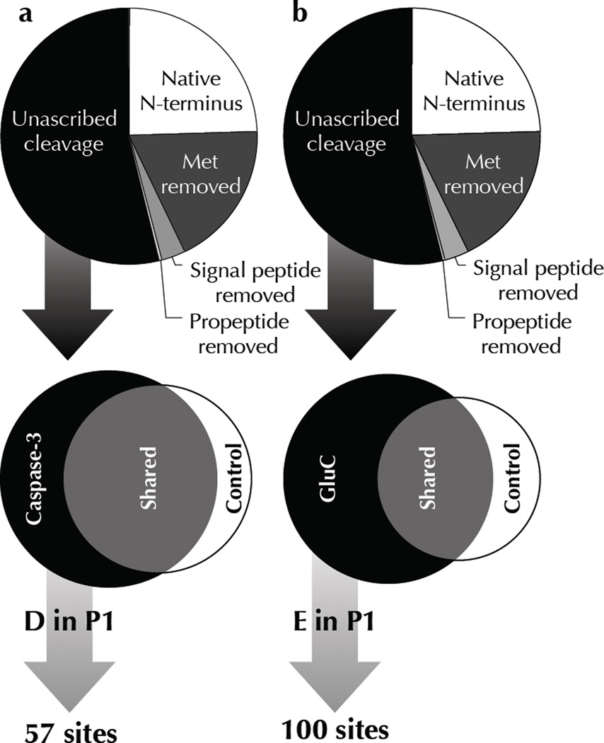
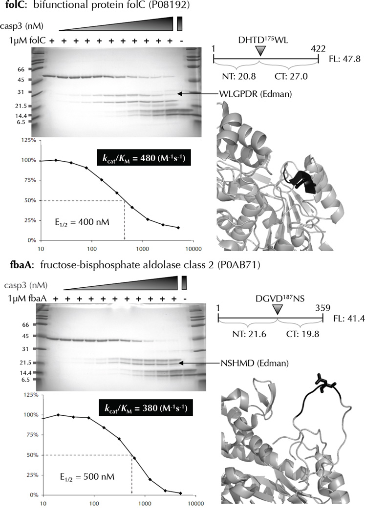
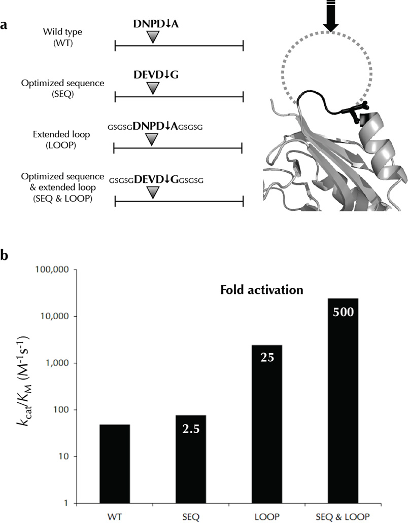
Two fundamental questions with regard to proteolytic networks and pathways concern the structural repertoire and kinetic threshold that distinguish legitimate signaling substrates. We used N-terminal proteomics to address these issues by identifying cleavage sites within the Escherichia coli proteome that are driven by the apoptotic signaling protease caspase-3 and the bacterial protease glutamyl endopeptidase (GluC). Defying the dogma that proteases cleave primarily in natively unstructured loops, we found that both caspase-3 and GluC cleave in alpha-helices nearly as frequently as in extended loops. Notably, biochemical and kinetic characterization revealed that E. coli caspase-3 substrates are greatly inferior to natural substrates, suggesting protease and substrate coevolution. Engineering an E. coli substrate to match natural catalytic rates defined a kinetic threshold that depicts a signaling event. This unique combination of proteomics, biochemistry, kinetics and substrate engineering reveals new insights into the structure-function relationship of protease targets and their validation from large-scale approaches.


Figures

Figure 1
Figure 1
Experimental approach. Samples of a library of structured proteins (E. coli soluble lysate) is treated with a range of exogenous protease concentrations (see text for details), and screened for cleavage-sites using N-terminomics. The location of cleavage sites is determined, and compared to available structures deposited in the Protein Data Bank, revealing the relationship between proteolysis and secondary structure.
Figure 2
Figure 2
N-terminomics reveals protease specific cleavage-sites. N-terminomic analysis of E. coli lysate treated with (a) human caspase-3 and (b) GluC reveals specific substrates and corresponding cleavage-sites. A tripartite criterion was used to identify genuine cleavage-sites of exogenous proteases from background proteolytic events inherent in E. coli lysate: Cleavage-sites must (1) not already be annotated as an endogenous site of proteolysis, (2) be only found in protease treated samples, and never in control samples, and (3) maintain the P1 aspartate/glutamate characteristic of each protease. See Table 1 for definitions.
Figure 3
Figure 3
Distribution of amino acids in the P1 position of unascribed cleavage-sites. Protease only cleavage-sites (black bars) reveal the hallmark P1 specificity of human caspase-3 (a) and GluC (b). Cleavage-sites found in control samples (grey bars) suggest endogenous proteolysis at Ala, Lys, and Arg residues in the P1 position, and confirm the lack of endogenous Asp or Glu specific protease activity in E. coli.
Figure 4
Figure 4
Specificity of caspase-3 and GluC. WebLogo representations of protease cleavage-sites depict the amino acid conservation and frequency at each position. The classic human caspase-3 consensus sequence DEVD↓G based on peptide positional scanning libraries , is recapitulated with two notable exceptions: there is no conservation at P3 and a weak additional preference for small uncharged amino acids at P2’ (a). GluC is not known to possess any extended specificity ; however, we see a weak preference for hydrophobic residues in the P1’ position (b).
Figure 5
Figure 5
Secondary structure preferences of human caspase-3 and GluC. WebLogo representations of secondary structures from protease cleavage-sites with the scissile bond indicated by the grey arrow. Secondary structure assignments were determined from (a,c) protein structures residing in the PDB as defined by DSSP, or (b,d) predicted by the PSIPRED algorithm. Both human caspase-3 and GluC cleaved substrates in loops as well as helices, but almost never in strands. The intolerance of human caspase-3 for β-strands appears to be restricted around the scissile bond. However, the strand intolerance for GluC is shifted toward the substrates N-terminus. Secondary structure assignments are as follows: L = loop, A = α-helix, B = β-strand.
Figure 6
Figure 6
Biochemical and kinetic analysis of cleavage-sites identified by N-terminomics. Many E. coli proteins that were identified as substrates of human caspase-3, and containing only 1 cleavage-site were recombinantly expressed, purified, and subjected to in vitro cleavage by human caspase-3. The cleavage-site P4-P1’ amino acids are colored black with the P1 residue in stick format. Substrate cleavage-sites were identified by N-terminal sequencing of the proteolytic fragments using Edman degradation. The E1/2 values were measured based on the disappearance of the full-length substrate using densitometry. These values were used to calculate kcat/KM for each substrate.
Figure 7
Figure 7
Engineered carA mutants are cleaved more efficiently than wild type. The E. coli caspase-3 substrate was engineered to dissect the contribution of sequence and structure on cleavage efficiency. The various constructs with are shown in (a) with an optimized sequence, an extended loop, and a combination of both. Relative rates of cleavage were measured for these mutants revealing that an extended loop conformation improves the kcat/KM more dramatically than an optimized sequence (b). However, an optimized sequence in parallel with an extended loop synergizes to elicit efficient cleavage.
Figure 8
Figure 8
Natural human caspase-3 substrates are kinetically superior to E. coli substrates. Human caspase-3 cleaves most E. coli substrates with kcat/KM between 50 and 2,000 M−1s−1, while several biologically relevant human caspase-3 substrates were cleaved with values greater than 10,000 M−1s−1. The propensity of natural substrates to be kinetically superior to E. coli substrates was shown to be statistically significant (p=0.0019). Engineering the E. coli substrate carA to contain an optimized cleavage-site sequence in an extended loop improved the kcat/KM value to over 30,000 M−1s−1. Some natural substrates have kcat/KM values greater than 30,000 M−1s−1, implying additional mechanisms for enhancing catalysis, discussed in the text.

References

    1. Puente XS, Sanchez LM, Overall CM, Lopez-Otin C. Human and mouse proteases: a comparative genomic approach. Nat Rev Genet. 2003;4:544–558. - PubMed
    1. Salvesen GS, Abrams JM. Caspase activation - stepping on the gas or releasing the brakes? Lessons from humans and flies. Oncogene. 2004;23:2774–2784. - PubMed
    1. Gevaert K, et al. Applications of diagonal chromatography for proteome-wide characterization of protein modifications and activity-based analyses. Febs J. 2007;274:6277–6289. - PubMed
    1. Timmer JC, Salvesen GS. Caspase substrates. Cell Death Differ. 2007;14:66–72. - PubMed
    1. Ding L, et al. Origins of the specificity of tissue-type plasminogen activator. Proc Natl Acad Sci USA. 1995;92:7627–7631. - PMC - PubMed

Publication types