

Inhibition of choroidal neovascularization by topical application of angiogenesis inhibitor vasostatin
- PMID: 19768130
- PMCID: PMC2746267
Inhibition of choroidal neovascularization by topical application of angiogenesis inhibitor vasostatin
Abstract
Purpose: Choroidal neovascularization (CNV) is the leading cause of blindness in patients with age-related macular degeneration (AMD). This study evaluated the inhibitory effect of vasostatin (VS), an endogenous angiogenesis inhibitor, on CNV.
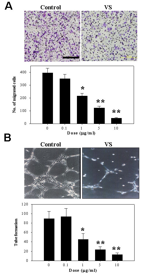
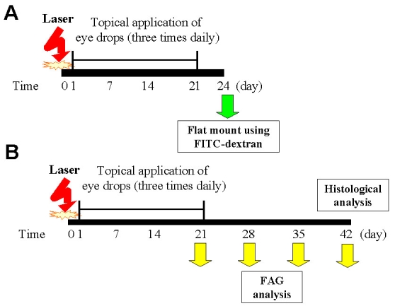
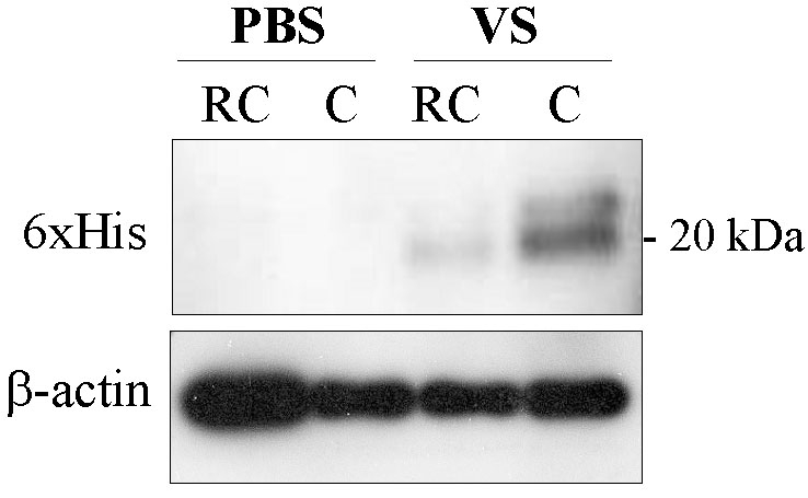
Methods: Anti-angiogenic activity of VS was evaluated in vitro by migration and tube formation assays in human umbilical vein endothelial cells (HUVECs). CNV lesions were induced in Brown Norway rats by fundus argon laser photocoagulation. Beginning one day after CNV induction, rats were treated with eye drops containing 1 microg/ml VS in PBS buffer for three times daily for 20 days. The extent of CNV was examined by flat mount analysis on day 24 or by fundus fluorescein angiography (FAG) on days 21, 28, 35, and 42, respectively. CNV lesions and choroidal vascularity were evaluated by histological analysis. The spatial distribution of topically applied VS in rat eyes was evaluated by immunoblot analysis.
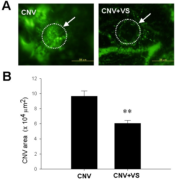
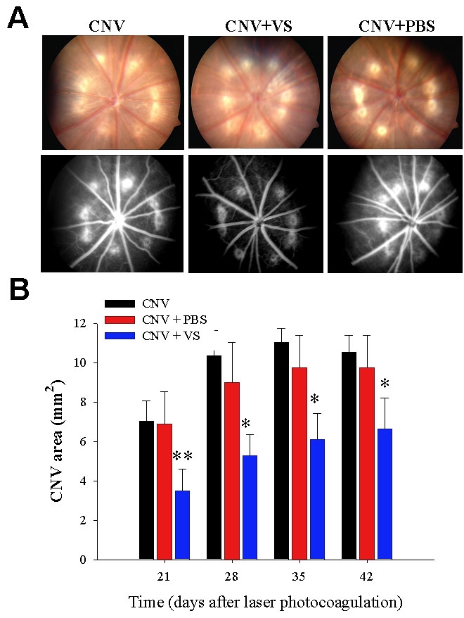
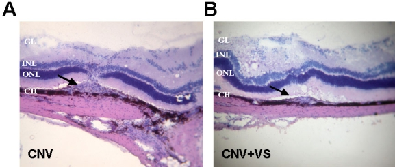
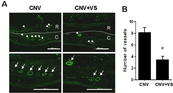
Results: VS inhibited migration and tube formation in HUVECs. Flat mount analysis revealed that, after laser-induced photocoagulation, topical VS application for 20 days significantly reduced CNV lesions. Moreover, serial FAG analysis indicated that a 20 day VS treatment significantly reduced CNV lesions on all subsequent days. Histological analysis revealed attenuated lesions, intact Bruch's membrane, and reduced choroidal vascularity in VS-treated eyes. Finally immunoblot analysis reveled VS expression in choroids.
Conclusions: Topical VS application suppresses the progression of laser-induced CNV via angiogenesis inhibition and may constitute a therapeutic alternative for excessive neovascularization occurring with ocular diseases.
Figures
Similar articles
-
Topical application of recombinant calreticulin peptide, vasostatin 48, alleviates laser-induced choroidal neovascularization in rats.Mol Vis. 2010 Apr 28;16:756-67. Mol Vis. 2010. PMID: 20454694 Free PMC article.
-
Inhibition of Experimental Choroidal Neovascularization by a Novel Peptide Derived from Calreticulin Anti-Angiogenic Domain.Int J Mol Sci. 2018 Sep 30;19(10):2993. doi: 10.3390/ijms19102993. Int J Mol Sci. 2018. PMID: 30274378 Free PMC article.
-
Suppression of choroidal neovascularization by intramuscular polymer-based gene delivery of vasostatin.Exp Eye Res. 2005 Dec;81(6):673-9. doi: 10.1016/j.exer.2005.04.005. Epub 2005 Jun 20. Exp Eye Res. 2005. PMID: 15967435
-
Inhibition of choroidal neovascularization by homoisoflavanone, a new angiogenesis inhibitor.Mol Vis. 2008 Mar 18;14:556-61. Mol Vis. 2008. PMID: 18385791 Free PMC article.
-
Effect of chromogranin A-derived vasostatin-1 on laser-induced choroidal neovascularization in the mouse.Acta Ophthalmol. 2015 May;93(3):e218-22. doi: 10.1111/aos.12557. Epub 2014 Oct 1. Acta Ophthalmol. 2015. PMID: 25271003
Cited by
-
Inducible expression of calreticulin-N58 in Pichia pastoris by high density cell culture.Mol Biol Rep. 2011 Nov;38(8):5003-8. doi: 10.1007/s11033-010-0646-5. Epub 2010 Dec 23. Mol Biol Rep. 2011. PMID: 21181274
-
Gene Therapy with Endogenous Inhibitors of Angiogenesis for Neovascular Age-Related Macular Degeneration: Beyond Anti-VEGF Therapy.J Ophthalmol. 2015;2015:201726. doi: 10.1155/2015/201726. Epub 2015 Mar 3. J Ophthalmol. 2015. PMID: 25821585 Free PMC article. Review.
-
Topical application of PPADS inhibits complement activation and choroidal neovascularization in a model of age-related macular degeneration.PLoS One. 2013 Oct 9;8(10):e76766. doi: 10.1371/journal.pone.0076766. eCollection 2013. PLoS One. 2013. PMID: 24130789 Free PMC article.
-
Topical application of recombinant calreticulin peptide, vasostatin 48, alleviates laser-induced choroidal neovascularization in rats.Mol Vis. 2010 Apr 28;16:756-67. Mol Vis. 2010. PMID: 20454694 Free PMC article.
-
Topical application of a G-Quartet aptamer targeting nucleolin attenuates choroidal neovascularization in a model of age-related macular degeneration.Exp Eye Res. 2015 Nov;140:171-178. doi: 10.1016/j.exer.2015.09.005. Epub 2015 Sep 12. Exp Eye Res. 2015. PMID: 26368850 Free PMC article.
References
-
- Campochiaro PA. Retinal and choroidal neovascularization. J Cell Physiol. 2000;184:301–10. - PubMed
-
- D'Amato RJ, Adamis AP. Angiogenesis inhibition in age-related macular degeneration. Ophthalmology. 1995;102:1261–2. - PubMed
-
- Campochiaro PA. Molecular targets for retinal vascular diseases. J Cell Physiol. 2007;210:575–81. - PubMed
-
- Gragoudas ES, Adamis AP, Cunningham ET, Jr, Feinsod M, Guyer DR. Pegaptanib for neovascular age-related macular degeneration. N Engl J Med. 2004;351:2805–16. - PubMed
-
- Avery RL, Pieramici DJ, Rabena MD, Castellarin AA, Nasir MA, Giust MJ. Intravitreal bevacizumab (Avastin) for neovascular age-related macular degeneration. Ophthalmology. 2006;113:363–72. - PubMed
Publication types
MeSH terms
Substances
LinkOut - more resources
Full Text Sources
Other Literature Sources