Review
. 2009:120:249-85.

Medical and genetic differences in the adverse impact of sleep loss on performance: ethical considerations for the medical profession

Affiliations
Review

Medical and genetic differences in the adverse impact of sleep loss on performance: ethical considerations for the medical profession

Charles A Czeisler. Trans Am Clin Climatol Assoc. 2009.

Abstract

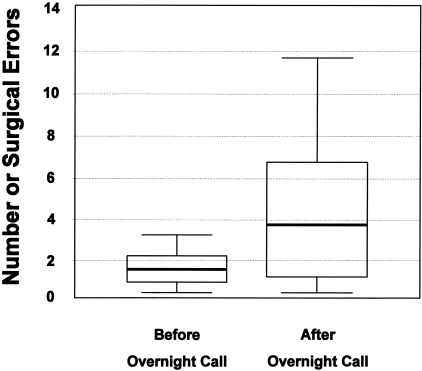
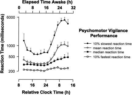
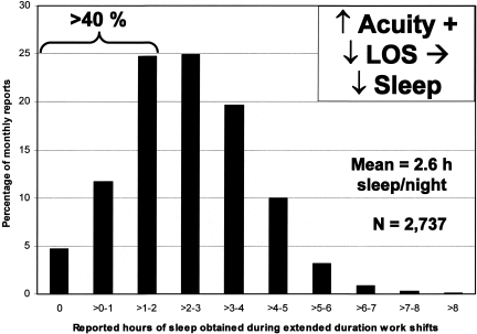
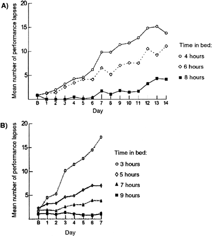
The Institute of Medicine recently concluded that-on average-medical residents make more serious medical errors and have more motor vehicle crashes when they are deprived of sleep. In the interest of public safety, society has required limitations on work hours in many other safety sensitive occupations, including transportation and nuclear power generation. Those who argue in favor of traditional extended duration resident work hours often suggest that there are inter- individual differences in response to acute sleep loss or chronic sleep deprivation, implying that physicians may be more resistant than the average person to the detrimental effects of sleep deprivation on performance, although there is no evidence that physicians are particularly resistant to such effects. Indeed, recent investigations have identified genetic polymorphisms that may convey a relative resistance to the effects of prolonged wakefulness on a subset of the healthy population, although there is no evidence that physicians are over-represented in this cohort. Conversely, there are also genetic polymorphisms, sleep disorders and other inter-individual differences that appear to convey an increased vulnerability to the performance-impairing effects of 24 hours of wakefulness. Given the magnitude of inter-individual differences in the effect of sleep loss on cognitive performance, and the sizeable proportion of the population affected by sleep disorders, hospitals face a number of ethical dilemmas. How should the work hours of physicians be limited to protect patient safety optimally? For example, some have argued that, in contrast to other professions, work schedules that repeatedly induce acute and chronic sleep loss are uniquely essential to the training of physicians. If evidence were to prove this premise to be correct, how should such training be ethically accomplished in the quartile of physicians and surgeons who are most vulnerable to the effects of sleep loss on performance without unacceptably compromising patient safety? Moreover, once it is possible to identify reliably those most vulnerable to the adverse effects of sleep loss on performance, will academic medical centers have an obligation to evaluate the proficiency of both residents and staff physicians under conditions of acute and chronic sleep deprivation? Should work-hour policy limits be modified to ensure that they are not hazardous for the patients of the most vulnerable quartile of physicians, or should the limits be personalized to enable the most resistant quartile to work longer hours? Given that the prevalence of sleep disorders has increased in our society overall, and increases markedly with age, how should fitness for extended duration work hours be monitored over a physician's career? In the spirit of the dictum to do no harm, advances in understanding the medical and genetic basis of inter-individual differences in the performance vulnerability to sleep loss should be incorporated into the development of work-hour policy limits for both physicians and surgeons.


Conflict of interest statement

Potential Conflicts of Interest: Dr. Czeisler is/was a consultant for: Actelion, Ltd.; Cephalon, Inc.; Delta Air Lines, Inc.; Eli Lilly and Co.; Garda Síochana Inspectorate; Global Ground Support; Johnson & Johnson; Koninklijke Philips Electronics, N.V.; Portland Trail Blazers; Respironics, Inc; Sanofi-Aventis Groupe; Sepracor, Inc.; Sleep Multimedia, Inc.; Somnus Therapeutics, Inc.; University of Wisconsin; Vanda Pharmaceuticals, Inc.; and Zeo, Inc.; and received royalties from McGraw Hill and Penguin Press. Dr. Czeisler owns an equity interest in Lifetrac, Inc.; Somnus Therapeutics, Inc.; Vanda Pharmaceuticals, Inc.; and Zeo, Inc. Dr. Czeisler has also received research support from Cephalon, Inc.; Tempur Pedic International, Inc; and Resmed, Inc. The Sleep and Health Education Program of the Harvard Medical School Division of Sleep Medicine has received support from Cephalon, Inc.; Takeda Pharmaceuticals North America, Inc.; Sanofi-Aventis Groupe; and Sepracor, Inc. Dr. Czeisler has received awards with monetary stipends from the American Clinical and Climatological Association; American Academy of Sleep Medicine; Association for Patient- Oriented Research; National Institute for Occupational Safety and Health and National Sleep Foundation; and the Sleep Research Society. Dr. Czeisler is the incumbent of an endowed professorship provided to Harvard University by Cephalon, Inc. and holds a number of process patents in the field of sleep/circadian rhythms (e.g., photic resetting of the human circadian pacemaker). Since 1985, Dr. Czeisler has also served as an expert witness on various legal cases related to sleep and/or circadian rhythms.

Figures

Fig. 1
Fig. 1
Effect of Partial Sleep Loss during Hospital Call on Errors Committed by Surgeons during Simulated Surgery. Simulated laparoscopic surgery performance of surgical residents (median age 34) measured by a laparoscopic surgery simulator (task 6 of the MIST-VR, Mentice Medical Simulation, Gothenburg, Sweden) before and after a night on call (17.5 hours from 3:30 pm to 9 am; median reported sleep time 1.5 h; range 0–3 h). Horizontal bands indicate median number of errors on this task, boxes show 25th and 75th centiles, and whisker lines show the highest and lowest error values. Figure and legend reprinted with permission from: Grantcharov TP, Bardram L, Funch-Jensen P, Rosenberg J. BMJ 2001;323:1222–1223 ().
Fig. 2
Fig. 2
Impact of Acute Total Sleep Deprivation on Reaction Time Performance. Time course of psychomotor vigilance task (PVT) performance [mean, median, 10% slowest and fastest reaction times in milliseconds (logarithmic scale)] during more than 28 hours of continuously monitored wakefulness under constant environmental and behavioral conditions are shown averaged across 10 subjects ± standard error of the mean. All data are binned from 10-minute PVT tests administered at 2-hour intervals and expressed with respect to elapsed time since wake time (designated as a Relative Clock Hour of 8), which was scheduled at its habitual hour. Figure and legend reprinted with permission from: Cajochen C, Khalsa SBS, Wyatt JK, Czeisler CA, Dijk D-J. Am J Physiol 277: R640–R649, 1999 ().
Fig. 3
Fig. 3
Sleep Duration in the Hospital. Average hours that interns reportedly slept in the hospital during extended duration (greater than 24 hour) on-call shifts as a percentage of 17,003 monthly survey reports collected from 2,737 resident physicians. Average hours of sleep obtained during extended duration shifts reported by interns for each week were averaged over the four weeks of each month to derive the value for each monthly survey report. Figure and legend reprinted with permission from: Barger LK, Cade BE, Ayas N, Cronin JW, Rosner B, Speizer FE, Czeisler CA, N Engl J Med 2005;352:125–134 ().
Fig. 4
Fig. 4
Repeated Nights of Sleep Loss Result in Cumulative Cognitive Impairment. Higher number of attentional performance failures on the PVT indicates poorer performance and more unstable alertness. Panel A shows the average number of lapses of attention recorded during 10- minute PVT tests administered eight times daily (every two hours) during 14 consecutive days when the amount of time in bed was limited to 4 hours, 6 hours or 8 hours per night. Lower panel shows the average number of lapses of attention recorded during 10-minute PVT tests administered four times daily during 7 consecutive days when the amount of time in bed was limited to 3 hours, 5 hours, 7 hours or 9 hours per night. Panel B signifies data from the baseline day. Figure and legend reprinted with permission from the 2006 IOM Report entitled: Sleep Deprivation and Sleep Disorders: An Unmet Public Health Problem ().
Fig. 5
Fig. 5
Subjective Sleepiness, Reaction Time, Lapses of Attention, and Attentional Failures Across 26 Hours of Wakefulness in Young and Older Participants. Group average data (+ standard error of the mean) are plotted with respect to time since scheduled awakening for 11 healthy older (mean age 68.1 ± 3.6 years; range 65 to 76 years; filled symbols) and 26 healthy young (mean age 21.9 ± 3.3 years; range 18 to 29 years; open symbols) adults. Dashed box indicates time of usual sleep episode. Subjective sleepiness ratings from the Karolinska Sleepiness Scale (KSS; scale range from 1=very alert to 9=very sleepy) are presented in Panel A. Mean reaction time (RT, in milliseconds) from each 10-minute PVT is presented in Panel B. As indicated in the first 16 hours of data, the RT on the PVT in well-rested individuals averages ∼250 milliseconds during the daytime, with minimal variability. The total number of lapses of attention (RT >500 milliseconds) from each 10-minute PVT are presented in Panel C. Well-rested individuals typically have very few (<5 per test administration) lapses of attention under these conditions. Attentional failures, defined as intrusions of slow eye movements (SEM) from continuous electro-oculographic recordings during EEG-verified wakefulness, were summed hourly and are presented in Panel D. Well-rested individuals typically have very few SEM under these conditions. Figure and legend reprinted with permission from: Duffy JD, Willson HJ, Wang W, Czeisler CA. J Am Geriatr Soc 2009; In Press. ().
Fig. 6
Fig. 6
Age Distribution of Crashes in Which Driver Was Not Intoxicated But Judged to Have Been Asleep. Data from 4,333 fall asleep crashes in North Carolina, where standard crash report forms, as mandated by state law, include ‘fatigued’ and ‘asleep’ in driver condition. Data for years 1990-1992, inclusive. In 55% of fall-asleep crashes, driver was age 25 or younger. Peak age of crashes was at age 20 years. Figure and legend reprinted with permission from: Pack AI, Pack AM, Rodgman E, Cucchiara A, Dinges DF, Schwab CW. Accid Anal Prev 27: 769, 1995 ().
Fig. 7
Fig. 7
Neurobehavioral responses to total sleep deprivation after two different prior sleep extension conditions. Each episode of sleep deprivation followed a week during which time the participants spent 12 hours in bed each night. The average number of lapses on 20-minute PVT tests administered every two hours over the last 24 hours of total sleep deprivation is shown. The abscissa shows the 19 individual participants, who were arbitrarily assigned the labels A through U. The participants are ordered by the magnitude of their impairment (averaged over the 2 sleep deprivations), with the most resistant participants on the left and the most vulnerable participants on the right. Responses in the first exposure to sleep deprivation following 7 days of prior sleep extension (mean ± standard deviation of the average nightly sleep duration of 8.5 ± 1.0 hours) are marked by boxes; responses in the second exposure to sleep deprivation following 7 days of prior sleep extension (mean ± standard deviation of the average nightly sleep duration of 8.8 ± 1.3 hours) are marked by diamonds. The panel reveals that subjects differed substantially in their responses to acute sleep deprivation, while the responses were relatively stable within subjects between the two exposures to acute sleep deprivation. Figure and legend reprinted with permission from: Van Dongen HPA, Baynard MD, Maislin G, Dinges DF. Sleep 2004; 27:423–433 ().
Fig. 8
Fig. 8
Polymorphism in Period Gene and Vulnerability to Sleep Loss. Left Hand Side: Deterioration of waking performance and increase of theta EEG activity and slow eye movements during sleep deprivation is greater in PER35/5 than in PER34/4 participants. Time course of central EEG theta (5–8 Hz) activity during wakefulness (Panel A), incidence of slow eye movement (SEMs) (percentage of 30 s epochs containing at least one SEM) (Panel B), and waking performance (composite performance score) (Panel C) are plotted relative to the timing of the plasma melatonin rhythm (Panel D) in ten PER35/5 (open symbols) and 14 PER34/4 (filled symbols) homozygotes. EEG theta activity, SEMs, and waking performance data were averaged per 2-hour intervals, relative to the midpoint of the melatonin rhythm. (* indicates a significant difference between genotypes, p < 0.05; upper abscissa indicates approximate wake duration.) Right Hand Side: Overnight performance on the paced visual serial addition task in PER35/5 and PER34/4 participants. Mean numbers of correct responses are plotted relative to the midpoint of the melatonin rhythm (Panel E). Overnight performance on the serial reaction time task in PER35/5 and PER34/4 participants. Mean switch costs, i.e., increase in time to respond to random rather than learned sequences of stimuli, are plotted relative to the melatonin midpoint. Higher values indicate poorer performance (Panel F). Overnight performance on spatial N-Back performance in relation to memory load in PER35/5 and PER34/4 participants. Mean numbers of correct responses, are plotted separately for the 1-, 2-, and 3-back, relative to the melatonin midpoint (Panel G). Overnight performance on verbal N-Back performance in relation to memory load in PER35/5 and PER34/4 participants. Mean numbers of correct responses are plotted separately for the 1-, 2-, and 3-back, relative to the melatonin midpoint (Panel H). * P < 0.05, Bonferroni corrected. Error bars represent the standard error of the mean. Figures and legends reprinted with permission from: Viola et al., Curr Biol, 2007 () (left hand side); and Groeger JA, Viola AU, Lo JC, von Schantz M, Archer SN, Dijk DJ. Sleep 2008;31:1159 () (right hand side).
Fig. 9
Fig. 9
Illustration of Eight Consecutive Days of a Typical Resident Physician Work Schedule Sanctioned by Current 2009 ACGME Guidelines. Every other shift is 30 hours in duration on this schedule. Because each 30-hour shift includes two calendar days, this schedule results in the resident physician spending every third night in the hospital. Black bar indicates scheduled 30-hour shifts. Hatched bar indicates an 8- to 10-hour swing shift, when resident physicians are not scheduled to work overnight. Box on right indicates how many hours residents are not scheduled to work for each 24-hour interval, beginning at 6 am each day.
Fig. 10
Fig. 10
.Reported Non-Compliance with ACGME Work-Hour Limits as Reported by Residents Non-Confidentially to the ACGME vs. Confidentially to the Harvard Work-Hours Health and Safety (HWHS) Group. After the ACGME implemented the 2003 limits on resident physicians work hours, 83.6% of interns reported to the HWHS Group work hours that were in violation of the ACGME standards during 1 or more months. Working shifts greater than 30 consecutive hours was reported by 67.4% of interns (open bar, middle pair). Averaged over 4 weeks, 43.0% of interns reported working more than 80 hours weekly (open bar, right pair), and 43.7% reported not having 1 day in 7 off work duties (data not shown). Violations were reported during 61.5% of months during which interns worked exclusively in inpatient settings. Violations were reported to the HWHS Group from 85.4% of the 707 represented residency programs (open bar, left pair). During the same reporting period, the ACGME reported near-universal compliance with the ACGME standards (filled bars), claiming that only 5.0% of residency training programs were not compliant with the standards and that only 3.3% of surveyed residents reported violations of the 80-hour rule. Differences between the ACGME and HWHS Group data were statistically significant on each of these measures (p < 0.001). Data and legend from: Landrigan CP, Barger LK, Cade BE, Ayas NT, Czeisler CA. JAMA 2006;296:1063 () Figure courtesy of Christopher P. Landrigan, M.D., M.P.H.

References

    1. Ulmer C, Wolman DM, Johns MME, editors. Resident Duty Hours: Enhancing Sleep, Supervision, and Safety. Washington, D.C.: The National Academies Press; 2008. Institute of Medicine of the National Academies; pp. 1–322. - PubMed
    1. Colten HR, Alteveogt BM, editors. Washington, D.C.: National Academies Press; 2006. Institute of Medicine. Sleep disorders and sleep deprivation: An unmet public health problem. . ISBN:0-309-66012-2. 1–500. - PubMed
    1. Assuring Quality Patient care and Quality Education. Association of American Medical Colleges; 2001. AAMC; pp. 1–10. - PubMed
    1. Friedman RC, Bigger JT, Kornfield DS. The intern and sleep loss. N Engl J Med. 1971;285:201–3. - PubMed
    1. Grantcharov TP, Bardram L, Funch-Jensen P, Rosenberg J. Laparoscopic performance after one night on call in a surgical department: prospective study. B M J. 2001;323:1222–3. - PMC - PubMed

Publication types

MeSH terms

Substances

LinkOut - more resources