. 2010 May 15;12(10):1147-54.
doi: 10.1089/ars.2009.2899.

The effect of hydrogen sulfide donors on lipopolysaccharide-induced formation of inflammatory mediators in macrophages

Affiliations

The effect of hydrogen sulfide donors on lipopolysaccharide-induced formation of inflammatory mediators in macrophages

Matthew Whiteman et al. Antioxid Redox Signal. .

Abstract

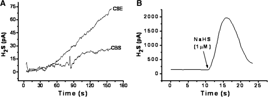
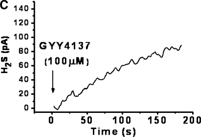
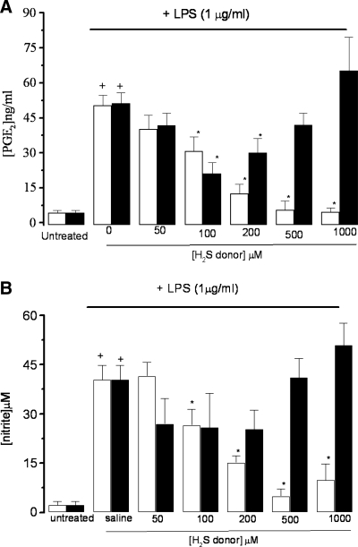
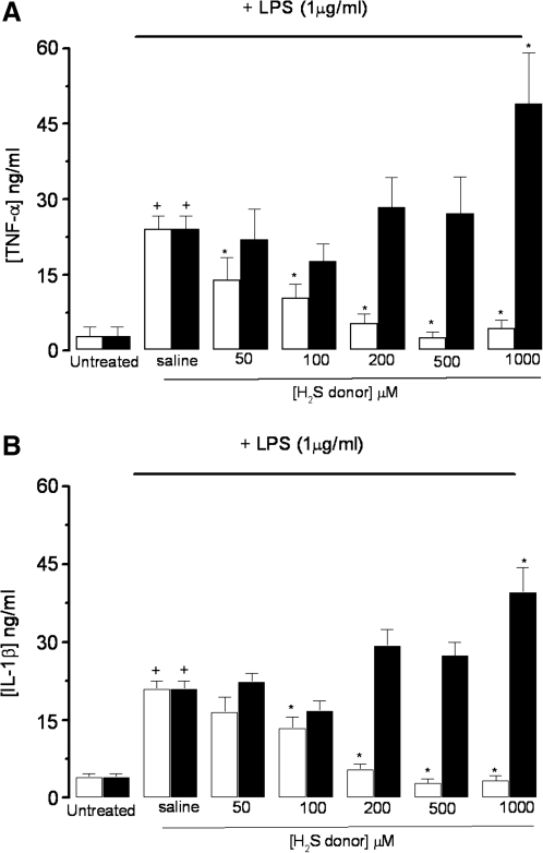
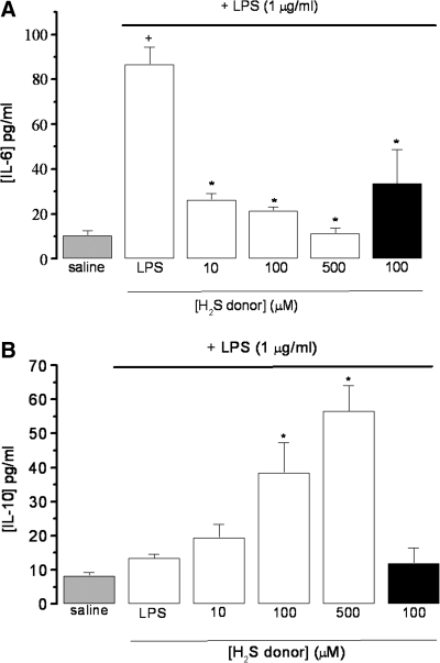
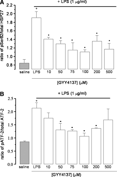
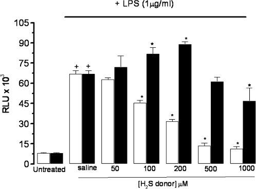
The role of hydrogen sulfide (H(2)S) in inflammation is controversial, with both pro- and antiinflammatory effects documented. Many studies have used simple sulfide salts as the source of H(2)S, which give a rapid bolus of H(2)S in aqueous solutions and thus do not accurately reflect the enzymatic generation of H(2)S. We therefore compared the effects of sodium hydrosulfide and a novel slow-releasing H(2)S donor (GYY4137) on the release of pro- and antiinflammatory mediators in lipopolysaccharide (LPS)-treated murine RAW264.7 macrophages. For the first time, we show that GYY4137 significantly and concentration-dependently inhibits LPS-induced release of proinflammatory mediators such as IL-1beta, IL-6, TNF-alpha, nitric oxide (*NO), and PGE(2) but increased the synthesis of the antiinflammatory chemokine IL-10 through NF-kappaB/ATF-2/HSP-27-dependent pathways. In contrast, NaHS elicited a biphasic effect on proinflammatory mediators and, at high concentrations, increased the synthesis of IL-1beta, IL-6, NO, PGE(2) and TNF-alpha. This study clearly shows that the effects of H(2)S on the inflammatory process are complex and dependent not only on H(2)S concentration but also on the rate of H(2)S generation. This study may also explain some of the apparent discrepancies in the literature regarding the pro- versus antiinflammatory role of H(2)S.


Figures

FIG. 1.
FIG. 1.
Time course of in vitro enzymatic formation of H2S from l-cysteine by recombinant CSE and CBS (A) and spontaneous H2S release from incubated NaHS (1 μM, B) and GYY4137 (100 μM, C). H2S was detected amperometrically. Results show representative traces from at least four separate experiments.
FIG. 1.
FIG. 1.
Time course of in vitro enzymatic formation of H2S from l-cysteine by recombinant CSE and CBS (A) and spontaneous H2S release from incubated NaHS (1 μM, B) and GYY4137 (100 μM, C). H2S was detected amperometrically. Results show representative traces from at least four separate experiments.
FIG. 2.
FIG. 2.
Effect of NaHS (black columns) and GYY4137 (open columns) on LPS (1 μg/ml)-evoked release of PGE2 (A) and nitrite (B) in incubated (24 h) RAW 264.7 cells. Results show concentration of PGE2 (ng/ml) or nitrite (micromolar) and are expressed as mean ± SEM; n = 5; *p < 0.05 (c.f. LPS group); +p < 0.05 (c.f. saline group); ANOVA plus post hoc Tukey test.
FIG. 3.
FIG. 3.
Effect of NaHS (black columns) and GYY4137 (open columns) on LPS (1 μg/ml)-evoked release of TNF-α (A) and IL-1β (B) in incubated (24 h) RAW 264.7 cells. Results show concentration of each cytokine (ng/ml) and are expressed as mean ± SE; n = 5; *p < 0.05 (c.f, LPS group); +p < 0.05 (c.f. saline group); ANOVA plus post hoc Tukey test.
FIG. 4.
FIG. 4.
Effect of NaHS (black column) and GYY4137 (open columns) on LPS (1 μg/ml)-evoked release of IL-6 (A) and IL-10 (B) in incubated (24 h) RAW 264.7 cells. Saline-treated control cells are shown by the grey column. Results show concentration of cytokines (pg/ml) and are expressed as mean ± SEM; n = 5–9; *p < 0.05 (c.f. LPS group); +p < 0.05 (c.f. saline group); ANOVA plus post hoc Tukey test.
FIG. 5.
FIG. 5.
Effect of GYY4137 on phosphorylation of ATF-2 (A) and HSP-27 (B) in LPS (1 μg/ml)-treated (24 h) RAW 264.7 cells. Saline-treated control cells are shown by the grey column. Results show ratio of phosphorylated to nonphosphorylated product and are expressed as mean ± SEM; n = 5; *p < 0.05 (c.f. LPS group); +p < 0.05 (c.f. saline group); ANOVA plus post hoc Tukey test.
FIG. 6.
FIG. 6.
Effect of NaHS (black columns) and GYY4137 (open columns) on activation of NF-κB in LPS (1 μg/ml)-treated (24 h) RAW 264.7 cells. Results show relative light units (RLU) and are expressed as mean ± SEM; n = 5; *p < 0.05 (c.f. LPS group); +p < 0.05 (c.f. saline group); ANOVA plus post hoc Tukey test.

References

    1. Alford KA. Glennie S. Turrell BR. Rawlinson L. Saklatvala J. Dean JL. Heat shock protein 27 functions in inflammatory gene expression and transforming growth factor-beta-activated kinase-1 (TAK1)-mediated signaling. J Biol Chem. 2007;282:6232–6241. - PubMed
    1. Bhatia M. Wong FL. Fu D. Lau HY. Moochhala SM. Moore PK. Role of hydrogen sulphide in acute pancreatitis in the mouse and rat. FASEB J. 2004;19:623–625. - PubMed
    1. Chang L. Geng B. Yu F. Zhao J. Jiang H. Du J. Tang C. Hydrogen sulfide inhibits myocardial injury induced by homocysteine in rats. Amino Acids. 2008;34:573–585. - PubMed
    1. Collin M. Anuar F. Murch O. Bhatia M. Moore PK. Thiemermann C. Inhibition of endogenous hydrogen sulfide formation reduces the organ injury caused by endotoxemia. Br J Pharmacol. 2005;146:498–505. - PMC - PubMed
    1. Florian B. Vintilescu R. Balseanu AT. Buga AM. Grisk O. Walker LC. Kessler C. Popa-Wagner A. Long-term hypothermia reduces infarct volume in aged rats after focal ischemia. Neurosci Lett. 2008;438:180–185. - PubMed

MeSH terms

LinkOut - more resources