Review
. 2009;13(5):221.
doi: 10.1186/cc7971. Epub 2009 Sep 8.

Bench-to-bedside review: Diaphragm muscle function in disuse and acute high-dose corticosteroid treatment

Affiliations
Review

Bench-to-bedside review: Diaphragm muscle function in disuse and acute high-dose corticosteroid treatment

Catherine Sh Sassoon et al. Crit Care. 2009.

Abstract

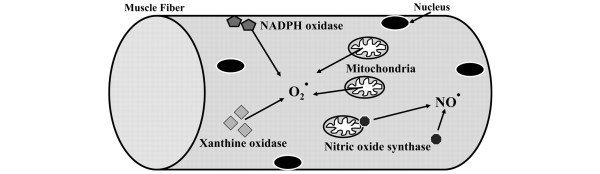
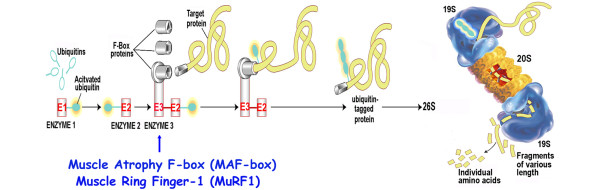
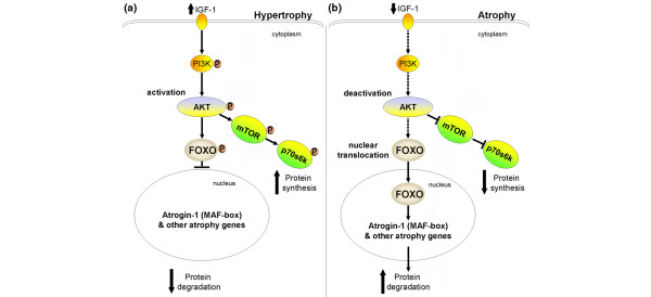
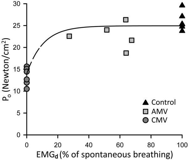
Critically ill patients may require mechanical ventilatory support and short-term high-dose corticosteroid to treat some specific underlying disease processes. Diaphragm muscle inactivity induced by controlled mechanical ventilation produces dramatic alterations in diaphragm muscle structure and significant losses in function. Although the exact mechanisms responsible for losses in diaphragm muscle function are still unknown, recent studies have highlighted the importance of proteolysis and oxidative stress. In experimental animals, short-term strategies that maintain partial diaphragm muscle neuromechanical activation mitigate diaphragmatic force loss. In animal models, studies on the influence of combined controlled mechanical ventilation and short-term high-dose methylprednisolone have given inconsistent results in regard to the effects on diaphragm muscle function. In the critically ill patient, further research is needed to establish the prevalence and mechanisms of ventilator-induced diaphragm muscle dysfunction, and the possible interaction between mechanical ventilation and the administration of high-dose corticosteroid. Until then, in caring for these patients, it is imperative to allow partial activation of the diaphragm, and to administer the lowest dose of corticosteroid for the shortest duration possible.


Figures

Figure 1
Figure 1
Oxidative stress pathways capable of producing reactive oxidant species. These pathways include nitric oxide synthase pathway, xanthine oxidase pathway, nicotinamide adenine dinucleotide phosphate (NADPH) oxidase pathway, and mitochondrial oxidant-generating pathway. The mitochondrial oxidant-generating pathway is key to oxidative damage of diaphragm muscle inactivity. O2+ superoxide; NO+, nitric oxide. Adapted with permission from [21].
Figure 2
Figure 2
The ubiquitin-proteasome pathway. The substrate proteins are designated for degradation by conjugation to ubiquitin in an ATP-dependent reaction. The ubiquitin-activating enzyme (E1) uses ATP to create a highly reactive thiolester form of ubiquitin, and then transfers it to a ubiquitin-carrier protein (E2). The subsequent transfer of the activated ubiquitin to the protein substrate requires a ubiquitin-protein ligase (E3). The E3 ligases muscle atrophy F-box (MAFbox) and muscle ring finger-1 (MuRF-1) have important roles in skeletal muscle atrophy. Once the ubiquitin conjugates are formed, they are transported to a proteolytic complex known as the 26S proteasome, consisting of two 19S regulators and the 20S core proteasome. The 19S regulators recognize and bind the ubiquitinated protein. Energy from ATP hydrolysis releases the ubiquitin chain and unfolds the substrate protein. The unfolded protein is fed into the 20S proteasome for degradation into small peptides and amino acids. The 20S proteasome can degrade oxidized protein without ubiquitination. Adapted with permission from [33].
Figure 3
Figure 3
The insulin-like growth factor-1-phosphotidylinositol-3-kinase-protein kinase-B serine/threonine kinase-forkhead box-O pathway. (a) Increased insulin-like growth factor-1 (IGF-1) activates phosphotidylinositol-3-kinase (PI3K), leading to phosphorylation of protein kinase-B serine/threonine kinase (Akt) and forkhead box-O (Foxo). Phosphorylated Foxo is sequestered within the cytoplasm and prevents its nuclear translocation and atrogin-1 (muscle atrophy F-box (MAFbox)) activation. Phosphorylated Akt also activates mammalian target of rapamycin (mTOR) and p70Sk, resulting in increased protein synthesis. (b) Suppression of IGF-1 with controlled mechanical ventilation-induced diaphragm muscle inactivity deactivates Akt, leading to nuclear translocation of Foxo, which then activates atrogin-1 and other atrogenes resulting in increased proteolysis. Reprinted from Cell, 117, Sandri M, Sandri C, Gilbert A, Skurk C, Calabria E, Picard A, Walsh K, Schiaffino S, Lecker SH, Goldberg AL, Foxo Transcription Factors Induce the Atrophy-Related Ubiquitin Ligase Atrogin-1 and Cause Skeletal Muscle Atrophy, 14 Pages, Copyright (2004), with permission from Elsevier [39].
Figure 4
Figure 4
Monoexponential relationships between diaphragm muscle maximal tetanic force and its electrical activity. The maximal isometric tension (Po) is normalized for muscle cross-sectional area. The diaphragm muscle electrical activity (EMGd) during assist-control mechanical ventilation (AMV) was estimated by measuring the area subtended by the moving average EMGd curve and its baseline, and is expressed as a percentage of spontaneous breathing. Po is maintained almost identically to that of the control after 3 days of AMV with diaphragm muscle activation between 30% and 80% of spontaneous breathing. Whether diaphragm muscle activation between 0% and 30% is effective to maintain Po remains unknown. Data obtained from [41]: n = 6 for the control and controlled mechanical ventilation (CMV) groups; n = 5 for the AMV group.
Figure 5
Figure 5
Cross-sectional areas of diaphragm muscle. Cross-sections of diaphragm muscle from biopsy specimens of a representative organ donor subject ((a), (c), (e)) and from a control ((b), (d), (f)). (a) and (b) Muscle fibers in the organ donor subject are in general smaller than those in the control diaphragm. No inflammatory infiltrate or necrosis is seen. Stained with hematoxylin and eosin. (c) and (d) Stained with antibody specific for slow myosin, heavy chain. (e) and (f) Stained with antibody specific for fast myosin, heavy chain. In (c) to (f), fibers reacting with the antibody appear orange-red, whereas fibers not reacting with the antibody appear black; open circle, slow-twitch fibers; open square, fast-twitch fibers. In addition, all fibers in each section are outlined by an antibody reactive to laminin. Reproduced with permission from [8]. Copyright © 2008 Massachusetts Medical Society. All rights reserved.

References

    1. Gainnier M, Roch A, Forel JM, Thirion X, Arnal JM, Donati S, Papazian L. Effect of neuromuscular blocking agents on gas exchange in patients presenting with acute respiratory distress syndrome. Crit Care Med. 2004;32:113–119. doi: 10.1097/01.CCM.0000104114.72614.BC. - DOI - PubMed
    1. Bracken MB, Shepard MJ, Holford TR, Leo-Summers L, Aldrich EF, Fazl M, Fehlings M, Herr DL, Hitchon PW, Marshall LF, Nockels RP, Pascale V, Perot PL Jr, Piepmeier J, Sonntag VK, Wagner F, Wilberger JE, Winn HR, Young W. Administration of methylprednisolone for 24 or 48 hours or tirilazad mesylate for 48 hours in the treatment of acute spinal cord injury. Results of the Third National Acute Spinal Cord Injury Randomized Controlled Trial. National Acute Spinal Cord Injury Study. JAMA. 1997;277:1597–1604. doi: 10.1001/jama.277.20.1597. - DOI - PubMed
    1. Nava S, Fracchia C, Callegari G, Ambrosino N, Barbarito N, Felicetti G. Weakness of respiratory and skeletal muscles after a short course of steroids in patients with acute lung rejection. Eur Respir J. 2002;20:497–499. doi: 10.1183/09031936.02.01732002. - DOI - PubMed
    1. Jagannath S. Treatment of myeloma in patients not eligible for transplantation. Curr Treat Options Oncol. 2005;6:241–253. doi: 10.1007/s11864-005-0007-0. - DOI - PubMed
    1. Kaplan PW, Rocha W, Sanders DB, D'Souza B, Spock A. Acute steroid-induced tetraplegia following status asthmaticus. Pediatrics. 1986;78:121–123. - PubMed

Publication types

MeSH terms

Substances