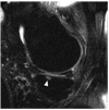
Subchondral bone marrow lesions are highly associated with, and predict subchondral bone attrition longitudinally: the MOST study
- PMID: 19769930
- PMCID: PMC2818146
- DOI: 10.1016/j.joca.2009.08.018
Subchondral bone marrow lesions are highly associated with, and predict subchondral bone attrition longitudinally: the MOST study
Abstract
Objective: Subchondral bone attrition (SBA) is defined as flattening or depression of the osseous articular surface. The causes of attrition are unknown, but remodeling processes due to chronic overload that are reflected as bone marrow edema-like lesions (BMLs) on magnetic resonance imaging (MRI) might predispose the subchondral bone to subsequent attrition. The aim of this study was to evaluate the cross-sectional and longitudinal association of BMLs with SBA in the same subregion of the knee.
Design: The Multicenter Osteoarthritis (MOST) study is a longitudinal observational study of individuals who have or are at high risk for knee osteoarthritis. Subjects with available baseline and 30-months follow-up MRI were included. Patients with a recent history of trauma or findings suggestive of post-traumatic bone marrow changes were excluded. Subchondral BMLs and SBA were scored semiquantitatively from 0 to 3 in 10 tibiofemoral subregions. We evaluated the association of prevalent BMLs at baseline with the presence of prevalent and incident SBA on a per-subregion basis using logistic regression. We also cross-sectionally evaluated the association of BML grade severity and presence of baseline SBA.
Results: One thousand and twenty-five knees were included. 8.9% of the analyzed knee subregions showed SBA present at baseline and 9.2% of subregions exhibited prevalent subchondral BMLs. The adjusted odds ratio (OR) for prevalent SBA for subregions with prevalent BMLs was 18.8 [95% confidence intervals (CI) 15.9-22.4]. A larger BML size was directly associated with an increased risk of prevalent SBA. 195 (2.2%) subregions exhibited incident SBA at follow-up. The adjusted OR for incident SBA was 5.3 [95% CI 3.6-7.7] when compared to subregions without BMLs as the reference.
Conclusions: Prevalent and incident SBA is strongly associated with subchondral BMLs in the same subregion. One explanation for the presence and development of SBA is subchondral remodeling due to increased stress, which is reflected as BMLs on MRI.
Copyright 2009 Osteoarthritis Research Society International. Published by Elsevier Ltd. All rights reserved.
Figures


References
-
- Arthritis prevalence and activity limitations--United States, 1990. MMWR Morb Mortal Wkly Rep. 1994;43:433–438. - PubMed
-
- Prevalence of disabilities and associated health conditions among adults--United States, 1999. MMWR Morb Mortal Wkly Rep. 2001;50:120–125. - PubMed
-
- Felson DT, McLaughlin S, Goggins J, LaValley MP, Gale ME, Totterman S, et al. Bone marrow edema and its relation to progression of knee osteoarthritis. Ann Intern Med. 2003;139:330–336. - PubMed
-
- Hunter DJ, Zhang Y, Niu J, Goggins J, Amin S, LaValley MP, et al. Increase in bone marrow lesions associated with cartilage loss: a longitudinal magnetic resonance imaging study of knee osteoarthritis. Arthritis Rheum. 2006;54:1529–1535. - PubMed
-
- Guermazi A, Zaim S, Taouli B, Miaux Y, Peterfy CG, Genant HG. MR findings in knee osteoarthritis. Eur Radiol. 2003;13:1370–1386. - PubMed
Publication types
MeSH terms
Grants and funding
LinkOut - more resources
Full Text Sources
Medical

