Comparative Study
. 2010 Feb 15;79(4):542-51.
doi: 10.1016/j.bcp.2009.09.017.

Withaferin A targets heat shock protein 90 in pancreatic cancer cells

Affiliations
Comparative Study

Withaferin A targets heat shock protein 90 in pancreatic cancer cells

Yanke Yu et al. Biochem Pharmacol. .

Abstract

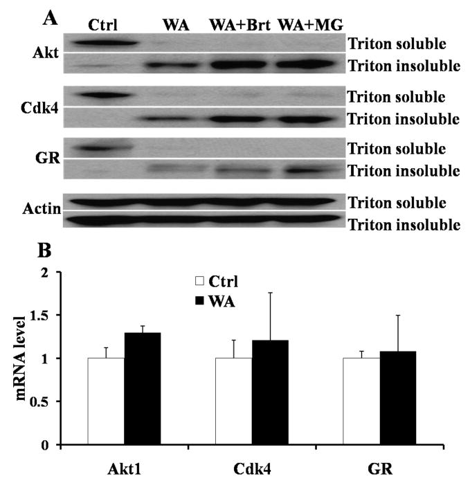
The purpose of this study is to investigate the efficacy and the mechanism of Hsp90 inhibition of Withaferin A (WA), a steroidal lactone occurring in Withania somnifera, in pancreatic cancer in vitro and in vivo. Withaferin A exhibited potent antiproliferative activity against pancreatic cancer cells in vitro (with IC(50)s of 1.24, 2.93 and 2.78 microM) in pancreatic cancer cell lines Panc-1, MiaPaCa2 and BxPc3, respectively. Annexin V staining showed that WA induced significant apoptosis in Panc-1 cells in a dose-dependent manner. Western blotting demonstrated that WA inhibited Hsp90 chaperone activity to induce degradation of Hsp90 client proteins (Akt, Cdk4 and glucocorticoid receptor), which was reversed by the proteasomal inhibitor, MG132. WA-biotin pull down assay of Hsp90 using Panc-1 cancer cell lysates and purified Hsp90 showed that WA-biotin binds to C-terminus of Hsp90 which was competitively blocked by unlabeled WA. Co-immunoprecipitation exhibited that WA (10 microM) disrupted Hsp90-Cdc37 complexes from 1 to 24h post-treatment, while it neither blocked ATP binding to Hsp90, nor changed Hsp90-P23 association. WA (3, 6mg/kg) inhibited tumor growth in pancreatic Panc-1 xenografts by 30% and 58%, respectively. These data demonstrate that Withaferin A binds Hsp90, inhibits Hsp90 chaperone activity through an ATP-independent mechanism, results in Hsp90 client protein degradation, and exhibits in vivo anticancer activity against pancreatic cancer.


Figures

Figure 1
Figure 1
A. Chemical structure of WA. B. Effect of WA treatment on viability of Panc-1 cells. Panc-1 cells were seeded in 96-well plates at a density of 5000 cells per well. 24 hours later the cells were subjected to WA treatment with concentrations of 0.05, 0.1, 0.5, 1.0, 2.5, 5, 10, and 20 μM. MTS assay was performed to assess cell viability after 48 h incubation. C. Effect of WA treatment on viability of MiaPaCa2 cells. Viability of MiaPaCa2 cells after WA treatment was assessed similar to B. D. Effect of WA treatment on viability of BxPc3 cells. Viability of BxPc3 cells after WA treatment was assessed similar to B.
Figure 2
Figure 2. WA induces apoptosis in Panc-1 cells as determined by Annexin V staining
A. Representative images from one of four independent experiments. Panc-1 cells were treated with 1, 5, and 10 μM WA for 12 h. Cells were stained with Annexin V-EGFP (green). Apoptotic cells were observed under fluorescent microscope. B. Quantification of WA induced apoptotic cells. The percentage of apoptotic cells were calculated as Annexin-EGFP positive cells divided by the total cancer cells.
Figure 3
Figure 3. Protein level changes after WA treatment in Panc-1 cells
A and B. WA induces Hsp90 client protein Akt and Cdk4 degradation in Panc-1 cells in a time- and dose-dependent manner. Panc-1 cells were treated with different concentrations of WA for different times. Cell lysates (50 μg protein in each lane) were analyzed by western blot analysis with specific antibodies to Akt, Cdk4 and Actin. Actin was served as internal standard. C. WA induces Hsp90 client protein GR degradation. Panc-1 cells were treated with10 μM WA for different times. Cell lysates (50 μg protein in each lane) were analyzed by western blot analysis with specific antibodies to GR and Actin. Actin was served as internal standard. D. WA induces expression of Hsp70. Panc-1 cells were treated with10 μM WA for different times. Cell lysates (50 μg protein in each lane) were analyzed by western blot analysis with specific antibodies to Hsp70 and Actin. Actin was served as internal standard. E. WA does not change the Cdk2 protein level. Panc-1 cells were treated with10 μM WA for different times. Cell lysates (50 μg protein in each lane) were analyzed by western blot analysis with specific antibodies to Cdk2 and Actin. Actin was served as internal standard.
Figure 4
Figure 4
A. WA-Biotin Hsp90 pull down assay.1 mg of Panc-1 cell lysates (Lysate), 5 μg of purified full length human Hsp90β (Full Hsp90) 5 μg of N-terminus human Hsp90β (N-Hsp90), 5 μg of C-terminus human Hsp90β (C-Hsp90) and 5 μg of yeast Hsp90 (Y-Hsp90) were used to carry out the WA-Biotin pull down assay. The WA-Biotin pull down protein were subjected to western blot analysis with specific antibodies to Hsp90. B. WA competes with WA-Biotin binding to Hsp90. The samples (Lysate, Full Hsp90 and C-Hsp90) were preincubated with 100 μM WA for 1 h before subject to WA-Biotin binding assay. The WA-Biotin pull down protein were subjected to western blot analysis with specific antibodies to Hsp90. C. WA induces Hsp90 aggregation in a dose dependent manner. Panc-1 cells were treated with 1, 5 and 10 μM WA for 24 h. Cell lysates (50 μg protein in each lane) were subjected to non-reducing gel electrophoresis and then analyzed by western blot with specific antibodies to Hsp90.
Figure 5
Figure 5
A. WA induced Hsp90 client protein degradation is proteasome-dependent. Panc-1 cells were preincubated with 10 μM Bortezomib (Brt) or MG132 (MG) for 1 h, and then were treated with 5 μM WA for another 12 h. Cells were collected and proteins were isolated as triton soluble part and triton insoluble part. Proteins (both triton soluble and triton insoluble parts) were subjected to western blot analysis with specific antibodies to Akt, Cdk4, GR and Actin. Actin was served as internal standard. B. mRNA levels of Hsp90 client protein in Panc-1 cells after WA treatment. Panc-1 cells were treated with 5 μM WA for 12 h, and the total mRNAs were isolated. RT-PCR was carried out to examine the mRNA levels of Akt, Cdk4 and GR using the specific primers of Akt, Cdk4, GR and Actin. Actin was served as internal standard.
Figure 6
Figure 6. WA does not inhibit ATP binding to Hsp90
5 μg of purified human Hsp90β was incubated with WA, 17-AAG and Celastrol (Cel) for 30 min. 25 μl ATP-sepharose beads were added to the samples to pull down Hsp90. Western blot was used to detect Hsp90 using specific antibody to Hsp90.
Figure 7
Figure 7. WA disrupts Hsp90-Cdc37 complex in Panc-1 cells
A and B. Co-immunoprecipitation (coIP) Hsp90. Cell lysates (500 μg total protein) were immunoprecipitated with Hsp90 antibody. Western blot was performed to detect Cdc37, Hop and Hsp90 using specific antibodies to Cdc37, Hop and Hsp90. A. Panc-1 cells were treated with 10 μM WA for 0 to 24 h. B. Panc-1 cells were treated with 1, 5, and 10 μM WA for 24 h. Input, total cell lysate; -IgG, without adding antibody. C. Western blot analysis of Cdc37 expression level. Panc-1 cells were treated with 10 μM WA for different times. Cell lysates (50 μg protein in each lane) were analyzed by western blot with specific antibodies to Cdc37 and Actin. Actin was served as internal standard. D. CoIP P23. Cell lysates (500 μg total protein) were immunoprecipitated with P23 antibody, then western blot was performed to detect Hsp90 and P23 using specific antibodies to Hsp90 and P23.
Figure 8
Figure 8. Antitumor effect of WA in Panc-1 xenografts
A. Tumor growth curves. The pancreatic tumor xenograft mouse model was generated by injecting the Panc-1 cancer cells s.c. to the right and left flanks of the nude mice. When the tumors reached 100 mm3, mice were divided randomly into three groups (n= 6/group) to receive vehicle, 3 mg/kg or 6 mg/kg WA treatment as scheduled. Tumor sizes and body weights were measured twice a week. Drug treatment was stopped after 30 days’ treatment, and tumor sizes and body weights were monitored until 70 days. Arrows indicate the date that stops treatment. B. Body weight of mice. Body weights of mice were monitored twice per week.

References

    1. Bardeesy N, DePinho RA. Pancreatic cancer biology and genetics. Nat Rev Cancer. 2002;2:897–909. - PubMed
    1. Jemal A, Siegel R, Ward E, Murray T, Xu J, Thun MJ. Cancer statistics. CA Cancer J Clin. 2007;57:43–66. - PubMed
    1. Di Costanzo F, Carlini P, Doni L, Massidda B, Mattioli R, Iop A, et al. Gemcitabine with or without continuous infusion 5-FU in advanced pancreatic cancer: a randomised phase II trial of the Italian oncology group for clinical research (GOIRC) British Journal of Cancer. 2005;93:185–9. - PMC - PubMed
    1. Kindler HL, Friberg G, Singh DA, Locker G, Nattam S, Kozloff M, et al. Phase II trial of bevacizumab plus gemcitabine in patients with advanced pancreatic cancer. J Clin Oncol. 2005;23:8033–40. - PubMed
    1. Xiong HQ, Rosenberg A, LoBuglio A, Schmidt W, Wolff RA, Deutsch J, et al. Cetuximab, a monoclonal antibody targeting the epidermal growth factor receptor, in combination with gemcitabine for advanced pancreatic cancer: a multicenter phase II Trial. J Clin Oncol. 2004;22:2610–6. - PubMed

Publication types

MeSH terms

LinkOut - more resources