. 2009 Dec 1;336(1):20-9.
doi: 10.1016/j.ydbio.2009.09.019. Epub 2009 Sep 19.

Absence of heartbeat in the Xenopus tropicalis mutation muzak is caused by a nonsense mutation in cardiac myosin myh6

Affiliations

Absence of heartbeat in the Xenopus tropicalis mutation muzak is caused by a nonsense mutation in cardiac myosin myh6

Anita Abu-Daya et al. Dev Biol. .

Abstract

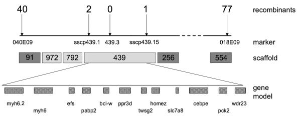
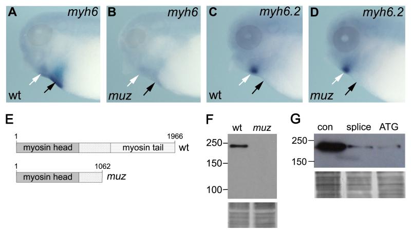
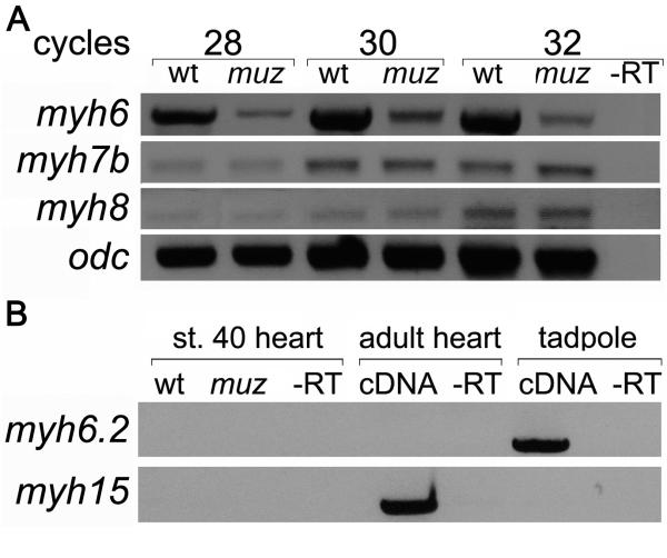
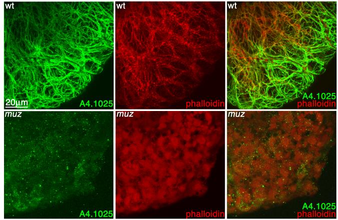
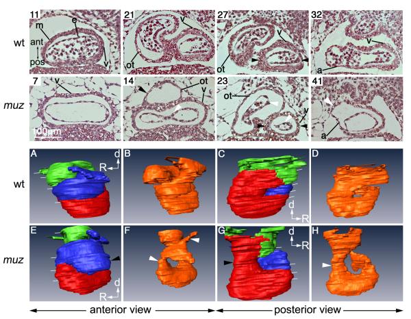
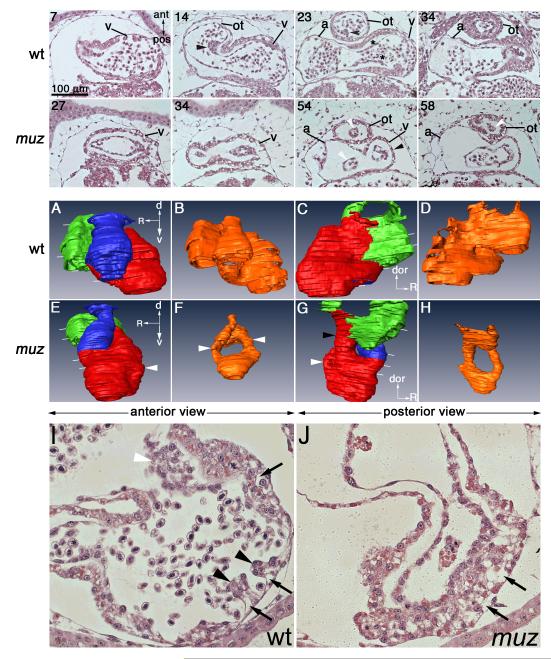
Mechanisms coupling heart function and cardiac morphogenesis can be accessed in lower vertebrate embryos that can survive to swimming tadpole stages on diffused oxygen. Forward genetic screens in Xenopus tropicalis have identified more than 80 mutations affecting diverse developmental processes, including cardiac morphogenesis and function. In the first positional cloning of a mutation in X. tropicalis, we show that non-contractile hearts in muzak (muz) embryos are caused by a premature stop codon in the cardiac myosin heavy chain gene myh6. The mutation deletes the coiled-coil domain responsible for polymerization into thick filaments, severely disrupting the cardiomyocyte cytoskeleton. Despite the lack of contractile activity and absence of a major structural protein, early stages of cardiac morphogenesis including looping and chamber formation are grossly normal. Muz hearts subsequently develop dilated chambers with compressed endocardium and fail to form identifiable cardiac valves and trabeculae.


Figures

Figure 1
Figure 1. Muz maps to an interval containing cardiac myosin heavy chain gene
A. Individual muz embryos were genotyped with SSLP markers from scaffold 91 and scaffold 554. Mapping was refined with SSCP markers (sscp439.1 and sscp439.15) and an SSLP marker (1.439.3) from scaffold 439; number of recombination events detected in 3200 meioses shown above each marker. Dark grey scaffolds are present on the tropicalis meiotic map; intervening light grey scaffolds were obtained by analysis of synteny to reference genomes and confirmed by linkage. Muz maps to a 370 kb genomic interval between sscp439.1 and sscp439.15 containing 12 gene models in the JGI assembly, including myh6 and myh6.2.
Figure 2
Figure 2. muzak is encoded by myh6
WISH shows myh6 expression in wild type heart (A, black arrow) and jaw muscle (white arrow) is diminished in muz (B). (C, D) myh6.2 is expressed in jaw muscle (white arrow) but not heart (black arrow), and is unaffected by the mutation. (E) Schematic showing domain structure of wild type X. tropicalis myh6 and the truncated protein lacking the myosin coiled-coil tail encoded by the muz allele. (F) Western blot analysis does not detect sarcomeric MHC protein in extracts of muz heart; silver stained loading control below. (Movie S2 and G) myh6 morphant hearts do not beat and show strong depletion of sarcomeric MHC protein relative to control morpholino-injected tadpoles; silver stained loading control below.
Figure 3
Figure 3. MHC genes expressed in stage 40 wild type and muz hearts
RT-PCR from isolated stage 40 hearts shows lower levels of myh6 in muz; myh7B and myh8 are unaffected. (A) myh6.2 mRNA is not detected in wild type or mutant tadpole hearts or wild type adult heart, although it is amplified from whole-embryo mRNA; myh15 is expressed in adult but not stage 40 tadpole heart(B).
Figure 4
Figure 4. Muz hearts lack myofibrils
3D confocal projections of wild type (A) and muz (B) hearts immunostained with the pan-sarcomeric MHC A4.1025 antibody (green) and counterstained with phalloidin (red). In wild type hearts, MHC and actin colocalize to myofibrils, while muz hearts show very little A4.1025 immunostaining and no fibrillar structures.
Figure 5
Figure 5. Altered chamber morphology in muz hearts
Coronal plastic sections of stage 40 wild type and muz hearts (top rows) numbered from ventral side of cardiac cavity, and indicated by white lines in 3D models (bottom rows). m= myocardium, e= inner endocardial tube, v= ventricle, ot= outflow tract , a= atrium. No blood cells are seen in the muz sections due to lack of circulation, and myocardial layer appears thinner throughout the muz heart compared to wild type. The muz ventricle is wider than in wild type (sections 7 and 11), while outflow tract and atrium are dilated (sections 14, 23 and 41). Abnormal muz chamber morphology is highlighted in 3D projections of outlines of myocardium (A, C, E, G, red=ventricle, blue=outflow tract, green=atrium) and endocardium (B, D, F, H, orange), including elongated ventricle, dilated outflow tract (black arrowhead in E) and narrow cardiac tube at AVC level (black arrow in G). muz endocardium is very compressed with drastically reduced lumen (white arrows in 23, F and H)
Figure 6
Figure 6. Muz hearts become dilated and lack valves and trabeculae
Coronal plastic sections of stage 42 wt and muz hearts (top rows) numbered from ventral side of cardiac cavity, and indicated by white lines in 3D models (middle rows). v= ventricle, ot= outflow tract , a= atrium. Wild type hearts show a spiral valve in the outflow tract (sections 14, 23, black arrows), and thickening of endocardium preceding atrioventricular valve formation (section 23, black asterisk). Valve formation is not detected in muz hearts, and endocardial lumen is drastically reduced in outflow tract and AVC regions (white arrowheads sections 54, 58, also compare models B and F). Endocardial cushion formation in AVC can also be seen in transverse sections of stage 42 wild type (I, white arrowhead) hearts but not in muz (J). Trabeculation has initiated in the wild type ventricle (I, black arrowheads) but is absent in muz (J). At this stage the ventricular myocardium has a vacuolated appearance in both wt and mutant embryos (I, J black arrows). Middle two rows: 3D projections of outlines of myocardium (A, C, E, G) and endocardium (B, D, F, H) highlight abnormal muz chamber morphology; red = ventricle, green = atrium, blue = outflow tract, orange = endocardium. Muz ventricles are elongated relative to wild type (E, G white arrows). A narrow tube connects muz ventricle and atrium (section 54 and G, black arrowheads; compare to 23, C).

References

    1. Armstrong EJ, Bischoff J. Heart valve development: endothelial cell signaling and differentiation. Circ Res. 2004;95:459–70. - PMC - PubMed
    1. Auman HJ, Coleman H, Riley HE, Olale F, Tsai HJ, Yelon D. Functional modulation of cardiac form through regionally confined cell shape changes. PLoS Biol. 2007;5:e53. - PMC - PubMed
    1. Bartman T, Walsh EC, Wen KK, McKane M, Ren J, Alexander J, Rubenstein PA, Stainier DY. Early myocardial function affects endocardial cushion development in zebrafish. PLoS Biol. 2004;2:E129. - PMC - PubMed
    1. Beis D, et al. Genetic and cellular analyses of zebrafish atrioventricular cushion and valve development. Development. 2005;132:4193–204. - PubMed
    1. Branford WW, Essner JJ, Yost HJ. Regulation of gut and heart left-right asymmetry by context-dependent interactions between xenopus lefty and BMP4 signaling. Dev Biol. 2000;223:291–306. - PubMed

Publication types

MeSH terms

LinkOut - more resources