Review
. 2009 Oct;30(6):713-43.
doi: 10.1210/er.2009-0005. Epub 2009 Sep 21.

Kisspeptin signaling in the brain

Affiliations
Review

Kisspeptin signaling in the brain

Amy E Oakley et al. Endocr Rev. 2009 Oct.

Abstract

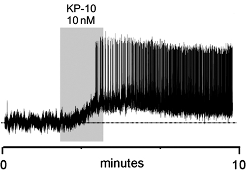
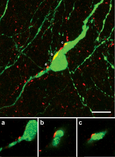
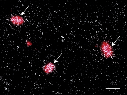
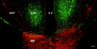
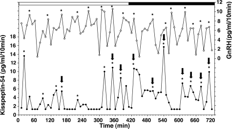
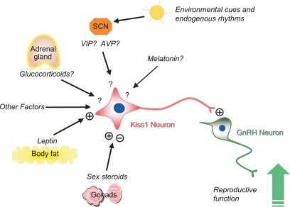
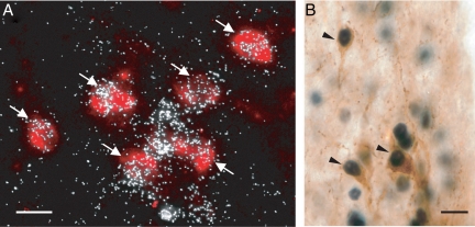
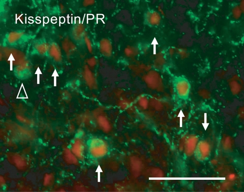
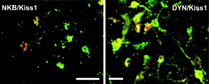
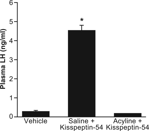
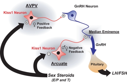
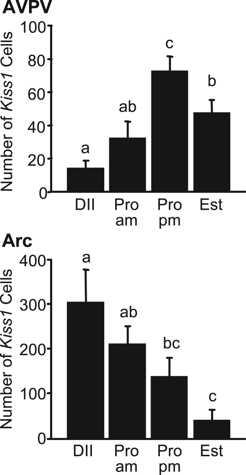
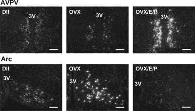
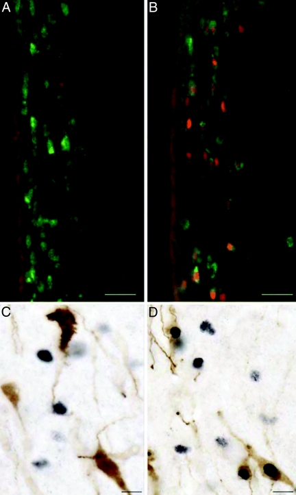
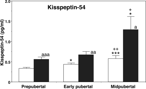
Kisspeptin (a product of the Kiss1 gene) and its receptor (GPR54 or Kiss1r) have emerged as key players in the regulation of reproduction. Mutations in humans or genetically targeted deletions in mice of either Kiss1 or Kiss1r cause profound hypogonadotropic hypogonadism. Neurons that express Kiss1/kisspeptin are found in discrete nuclei in the hypothalamus, as well as other brain regions in many vertebrates, and their distribution, regulation, and function varies widely across species. Kisspeptin neurons directly innervate and stimulate GnRH neurons, which are the final common pathway through which the brain regulates reproduction. Kisspeptin neurons are sexually differentiated with respect to cell number and transcriptional activity in certain brain nuclei, and some kisspeptin neurons express other cotransmitters, including dynorphin and neurokinin B (whose physiological significance is unknown). Kisspeptin neurons express the estrogen receptor and the androgen receptor, and these cells are direct targets for the action of gonadal steroids in both male and female animals. Kisspeptin signaling in the brain has been implicated in mediating the negative feedback action of sex steroids on gonadotropin secretion, generating the preovulatory GnRH/LH surge, triggering and guiding the tempo of sexual maturation at puberty, controlling seasonal reproduction, and restraining reproductive activity during lactation. Kisspeptin signaling may also serve diverse functions outside of the classical realm of reproductive neuroendocrinology, including the regulation of metastasis in certain cancers, vascular dynamics, placental physiology, and perhaps even higher-order brain function.


Figures

Figure 1
Figure 1
Proposed mechanism of neuronal depolarization by kisspeptin binding to its receptor, Kiss1r. Kisspeptin binding to its GPCR, Kiss1r, activates the G protein, Gαq, and PLC to cleave phosphatidylinositol 4,5-bisphosphate (PIP2) into IP3 and DAG. DAG activates a signal cascade by activating PKC, whereas IP3 mobilizes calcium ions (Ca2+), which participate in the cascade by activating other proteins. Membrane depolarization is caused by activation (+) of nonselective TRPC cation channels and inhibition (−) of inwardly rectifying potassium channels (Kir), possibly through involvement of DAG.
Figure 2
Figure 2
Products of the Kiss1 gene. Kiss1 mRNA is transcribed from the Kiss1 gene and translated to form a 145-amino-acid propeptide called kisspeptin-145. Shown are cleavage sites on the propeptide that lead to the production of the RF-amidated kisspeptin-54. Shorter peptides (such as kisspeptin-10, -13, and -14) share a common C terminus and RF-amidated motif with kisspeptin-54. Because no putative cleavage sites have been identified on the propeptide that would lead to synthesis of the shorter peptides, such peptides may be degradation products of kisspeptin-54. [Adapted with permission from Popa et al., 2008 (202) © Annual Reviews].
Figure 3
Figure 3
Effect of POA infusion of antirat kisspeptin monoclonal antibody on the proestrous LH surge. PBS (n = 6) or antirat kisspeptin monoclonal antibody (n = 5) was infused into the POA at 1000–1800 h. Blood samples were collected every hour at 1300–2000 h through an indwelling atrial cannula. Values are means ± sem. *, P < 0.05 vs. vehicle-treated control (one-way ANOVA with repeated measures). [Modified with permission from Kinoshita et al., 2005 (42) © The Endocrine Society].
Figure 4
Figure 4
Kisspeptin-10 sequences. Alignment of deduced amino acid sequences (from BLAST) of Kiss1 and Kiss2 in vertebrates. Conserved amino acid residues are in bold.
Figure 5
Figure 5
Kisspeptin exerts a potent activational effect on GnRH neurons in adult female proestrous mice. Perforated-patch, voltage recordings from proestrous female GnRH-GFP neurons (resting membrane potential = −68 mV) in the acute brain slice demonstrate a remarkably intense and prolonged activation by 10 nm kisspeptin (KP-10). [Modified with permission from Han et al., 2005 (70)].
Figure 6
Figure 6
Kisspeptin projections to GnRH neurons in adult female mice. Confocal stack of 75 images showing a single GnRH neuron (green) with kisspeptin (red) fibers surrounding and apposed to it. Single 370-nm-thick optical sections through the three regions indicated by a, b, and c of the GnRH neuron are given below to demonstrate the close apposition between kisspeptin fibers and GnRH neuron elements. Scale bar, 10 μm. [Modified with permission from Clarkson and Herbison, 2006 (73) © The Endocrine Society].
Figure 7
Figure 7
Coexpression of GnRH mRNA with Kiss1 mRNA in rats. Representative photomicrograph from the medial preoptic area. GnRH-mRNA-expressing cells are fluorescent with Vector Red substrate. Clusters of silver grains (white dots) reflect the presence of Kiss1r mRNA. The arrows indicate GnRH neurons that coexpress Kiss1r. Approximately 77% of the GnRH neurons coexpress Kiss1r mRNA (n = 4 rats). Scale bar, 20 μm. [Reproduced with permission from Irwig et al., 2004 (80) © S. Karger AG].
Figure 8
Figure 8
Structural interactions between Kiss1 and GnRH neurons in the mediobasal hypothalamus of the male rhesus monkey. A confocal projection (×10; 1-μm optical sections) illustrating the distribution of kisspeptin neurons (green fluorescence, Alexa Fluor 488) in relation to the GnRH neuronal network (red fluorescence, Cy3) in a coronal section of the MBH of an agonadal male rhesus monkey aged 4 yr 3 months. Whereas kisspeptin perikarya were confined to the ARC, those of GnRH extended along the ventral hypothalamic tract (VHT), lateral to the ARC. GnRH innervation of the external zone of the ME was intense. Beaded kisspeptin axons projected to the ME, and at this anteroposterior level GnRH and kisspeptin fibers running in a near horizontal plane were found in close association. 3V, Third ventricle. Scale bar, 100 μm. [Modified with permission from Ramaswamy et al., 2008 (99) © The Endocrine Society].
Figure 9
Figure 9
Kisspeptin-54 release in the stalk-ME of the monkey, as assessed by microdialysis, is pulsatile. Both kisspeptin-54 (closed circle) and GnRH (open circle) were measured in the same microdialysate samples. Kisspeptin-54 and GnRH pulses, indicated by asterisks, were identified using the PULSAR algorithm. Kisspeptin-54 pulses correlated with GnRH pulses are indicated by arrows on the top of kisspeptin pulses. Lighting conditions are indicated on the top of each graph (white bar for the lights-on period and black bar for the lights-off period). [Modified with permission from Keen et al., 2008 (89) © The Endocrine Society].
Figure 10
Figure 10
Kisspeptin neurons may act as central processors for relaying signals from the periphery to GnRH neurons. Metabolic and environmental factors regulate reproductive function, which ensures that reproduction proceeds only when metabolic and environmental conditions are favorable. Kisspeptin stimulates GnRH secretion, and Kiss1 mRNA is both negatively and positively regulated by sex steroids. The expression of Kiss1 may be induced by leptin, whose plasma levels reflect the state of metabolic reserves. Kisspeptin neurons may also receive input from the hypothalamic-pituitary-adrenal axis and from environmental cues such as time of day via the SCN of the hypothalamus and day length via melatonin from the pineal gland. AVP, Arginine vasopressin; VIP, vasoactive intestinal peptide. [Modified from Dungan et al., 2006 (203)].
Figure 11
Figure 11
Kisspeptin neurons express ERα. A, Representative photomicrograph showing coexpression of Kiss1 mRNA with ERα in the AVPV of the female mouse. Kiss1 mRNA-expressing cells were visualized with Vector Red substrate, and ERα was marked by the presence of clusters of silver grains (white dots). Arrows indicate Kiss1 neurons that coexpress ERα. Scale bar, 20 μm. B, Dual-label immunocytochemistry showing kisspeptin neurons (brown) with ERα-immunoreactive nuclei (black) in the AVPV of the female mouse. Arrowheads indicate dual-labeled cells. Scale bar, 10 μm. [Modified with permission from Smith et al., 2005 (104), © The Endocrine Society, and Clarkson et al., 2008 (106)].
Figure 12
Figure 12
Expression of PR in Kiss1 neurons. Photomicrograph of cells costaining for kisspeptin (green) and PR (red) in the ARC of the ewe brain. Arrows indicate cells containing both kisspeptin and PR. The open arrowhead indicates a kisspeptin-positive PR-negative cell. Scale bar, 50 μm. [Modified with permission from Smith et al., 2007 (107) © The Endocrine Society].
Figure 13
Figure 13
Coexpression of NKB/Kiss1 and dynorphin (DYN)/Kiss1 in the middle ARC of the ewe. Overlay of red (Kiss1 neurons) and green (NKB) fluorescent images (left; scale bar, 50 μm) and overlay of red (DYN) and green (Kiss1 neurons) fluorescent images (right; scale bar, 20 μm), illustrating colocalization of kisspeptin with NKB and DYN with kisspeptin, respectively. Double-labeling appears brown. [Modified with permission from Goodman et al., 2007 (110) © The Endocrine Society].
Figure 14
Figure 14
The GnRH antagonist, acyline, blocks the effects of kisspeptin-54 on plasma LH in the mouse. Kisspeptin-54 (50 pmol) and vehicle were administered into the lateral ventricle. Pretreatment with the GnRH antagonist, acyline (50 μg) or saline was given sc. *, P < 0.001 saline + kisspeptin-54 vs. all other treatments. [Modified with permission from Gottsch et al., 2004 (72) © The Endocrine Society].
Figure 15
Figure 15
A schematic representation of our current understanding of Kiss1 signaling in the forebrain of the mouse. Kisspeptin stimulates GnRH secretion by a direct effect on GnRH neurons, most of which express the kisspeptin receptor, Kiss1r. Neurons that express Kiss1 mRNA reside in the AVPV and the ARC (arcuate). Kiss1 neurons in the ARC appear to be involved in the negative feedback regulation of GnRH/LH by sex steroids. The expression of Kiss1 mRNA in the arcuate is inhibited by estradiol (E), progesterone (P), and testosterone (T). These same hormones induce Kiss1 mRNA expression in the AVPV, where Kiss1 neurons are thought to be involved in the positive feedback regulation of GnRH/LH. [Modified with permission from Gottsch et al., 2006 (204) © Elsevier].
Figure 16
Figure 16
Kiss1 expression in the AVPV and ARC over the estrous cycle of the rat. Values without common notations (a, b, c) differ significantly (P < 0.05). Values are the mean ± sem. DII, Diestrus; Pro am, proestrus 2 h after lights on; Pro pm, proestrus 1 h before lights off; Est, estrus. [From Smith et al., 2006 (82)].
Figure 17
Figure 17
Dark-field photomicrographs showing Kiss1 mRNA-expressing cells (as reflected by the presence of white clusters of silver grains) in representative sections of the AVPV and ARC from diestrus (DII), ovariectomized (OVX) and ovariectomized/estradiol/progesterone (OVX/E/P)-treated female rats. 3V, Third ventricle. Scale bars, 100 μm. [From Smith et al., 2006 (82)].
Figure 18
Figure 18
Fos expression in the AVPV of the rat before and during an LH surge. A and B, Photomicrographs showing Kiss1 neurons (green) in the AVPV on diestrus II (A) and proestrus (B), with the induction of Fos (red) in Kiss1 neurons on the afternoon of proestrus (compare A and B). Scale bars, 25 μm. C and D, Photomicrographs showing GnRH neurons (brown) and Fos (black) on diestrus II (C) and their coexpression on the afternoon of proestrus (D). Scale bars, 10 μm. [From Smith et al., 2006 (82)].
Figure 19
Figure 19
Developmental changes in the release of kisspeptin-54 in push-pull perfusates in monkeys. Kisspeptin-54 levels gradually increase along with the pubertal increase in GnRH release. A nocturnal increase in kisspeptin-54 release is already observed in prepubertal monkeys and continues through the pubertal period. The number of animals at the prepubertal, early pubertal, and midpubertal stages was six, six, and five, respectively. White bars, Morning values; black bars, evening values. *, P < 0.05 vs. prepubertal; ***, P < 0.001 vs. prepubertal; +, P < 0.05 vs. early pubertal; ++, P < 0.01 vs. early pubertal; a, P < 0.05 vs. morning; aa, P < 0.01 vs. morning; aaa, P < 0.001 vs. morning. [Modified with permission from Keen et al., 2008 (89) © The Endocrine Society].

References

    1. Lee JH, Miele ME, Hicks DJ, Phillips KK, Trent JM, Weissman BE, Welch DR 1996 KiSS-1, a novel human malignant melanoma metastasis-suppressor gene. J Natl Cancer Inst 88:1731–1737 - PubMed
    1. Kotani M, Detheux M, Vandenbogaerde A, Communi D, Vanderwinden JM, Le Poul E, Brézillon S, Tyldesley R, Suarez-Huerta N, Vandeput F, Blanpain C, Schiffmann SN, Vassart G, Parmentier M 2001 The metastasis suppressor gene KiSS-1 encodes kisspeptins, the natural ligands of the orphan G protein-coupled receptor GPR54. J Biol Chem 276:34631–34636 - PubMed
    1. Muir AI, Chamberlain L, Elshourbagy NA, Michalovich D, Moore DJ, Calamari A, Szekeres PG, Sarau HM, Chambers JK, Murdock P, Steplewski K, Shabon U, Miller JE, Middleton SE, Darker JG, Larminie CG, Wilson S, Bergsma DJ, Emson P, Faull R, Philpott KL, Harrison DC 2001 AXOR12, a novel human G protein-coupled receptor, activated by the peptide KiSS-1. J Biol Chem 276:28969–28975 - PubMed
    1. Ohtaki T, Shintani Y, Honda S, Matsumoto H, Hori A, Kanehashi K, Terao Y, Kumano S, Takatsu Y, Masuda Y, Ishibashi Y, Watanabe T, Asada M, Yamada T, Suenaga M, Kitada C, Usuki S, Kurokawa T, Onda H, Nishimura O, Fujino M 2001 Metastasis suppressor gene KiSS-1 encodes peptide ligand of a G-protein-coupled receptor. Nature 411:613–617 - PubMed
    1. Clements MK, McDonald TP, Wang R, Xie G, O'Dowd BF, George SR, Austin CP, Liu Q 2001 FMRFamide-related neuropeptides are agonists of the orphan G-protein-coupled receptor GPR54. Biochem Biophys Res Commun 284:1189–1193 - PubMed