. 2009 Oct;119(10):3070-8.
doi: 10.1172/JCI39269. Epub 2009 Sep 21.

Evidence of premature immune aging in patients thymectomized during early childhood

Affiliations

Evidence of premature immune aging in patients thymectomized during early childhood

Delphine Sauce et al. J Clin Invest. 2009 Oct.

Abstract

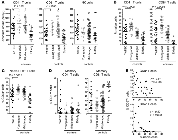
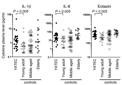
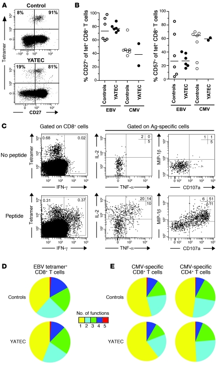
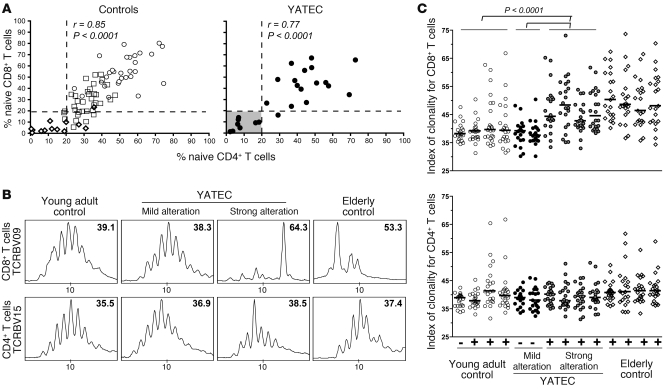
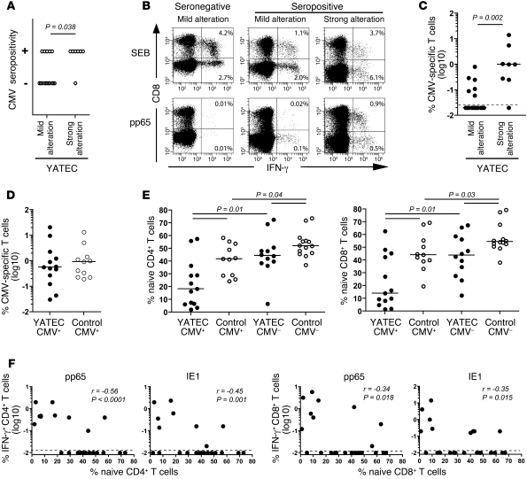
While the thymus is known to be essential for the initial production of T cells during early life, its contribution to immune development remains a matter of debate. In fact, during cardiac surgery in newborns, the thymus is completely resected to enable better access to the heart to correct congenital heart defects, suggesting that it may be dispensable during childhood and adulthood. Here, we show that young adults thymectomized during early childhood exhibit an altered T cell compartment. Specifically, absolute CD4+ and CD8+ T cell counts were decreased, and these T cell populations showed substantial loss of naive cells and accumulation of oligoclonal memory cells. A subgroup of these young patients (22 years old) exhibited a particularly altered T cell profile that is usually seen in elderly individuals (more than 75 years old). This condition was directly related to CMV infection and the induction of strong CMV-specific T cell responses, which may exhaust the naive T cell pool in the absence of adequate T cell renewal from the thymus. Together, these marked immunological alterations are reminiscent of the immune risk phenotype, which is defined by a cluster of immune markers predictive of increased mortality in the elderly. Overall, our data highlight the importance of the thymus in maintaining the integrity of T cell immunity during adult life.


Figures

Figure 1
Figure 1. Durable reduction of thymic mass after early thymectomy.
Representative CT scans of the heart region in a 5-year-old child thymectomized within 2 weeks of birth and a 5-year-old control subject. Axial and sagittal sections are shown. The arrows indicate the thymus area.
Figure 2
Figure 2. T cell count and subset distribution in thymectomized adults.
(A) Absolute counts for CD4+, CD8+ T cells and NK cells in YATECs (median age, 22.0 years). Counts in young adult (median age, 21.9 years), middle-aged (median age, 35.3 years), and elderly (median age, 82 years) controls are shown for comparison. (B) Proportions of naive (CD45RA+CCR7+CD27+) CD4+ or CD8+ T cells in YATECs and young, middle-aged, or elderly controls. (C) Proportions of CD31+ naive CD4+ T cells. (D) Proportions of CD57+ cells within memory CD4+ or CD8+ T cells. (E) Correlation between naive and CD57+ memory CD4+ or CD8+ T cell percentages in YATECs. The horizontal lines in AD indicate the median. P values were calculated by the Mann-Whitney U test for group comparisons. Spearman’s rank test was used to determine correlations.
Figure 3
Figure 3. Proinflammatory cytokine profile in YATECs.
Plasma levels of the proinflammatory cytokines IL-1β, IL-8, and eotaxin in YATECs, and young adult, middle-aged, or elderly control subjects. The horizontal lines indicate the median. P values were calculated using the Mann-Whitney U test for group comparisons.
Figure 4
Figure 4. Phenotypic and functional assessment of virus-specific CD8+ T cells in YATECs.
(A) Representative staining of CD27 on HLA-A11 EBV EBNA-3B AK10–specific CD8+ T cells identified in 1 YATEC and 1 control subject. Percentages of CD27 or CD27+ cells within tetramer-positive populations are shown. (B) Expression of the differentiation markers CD27 or CD57 on EBV- or CMV-specific CD8+ T cells, identified using tetramers in YATECs or control individuals. Horizontal bars indicate the median. (C) Representative example of simultaneous multifunctional assessment of B8-FL9 EBV–specific CD8+ T cells from a YATEC patient. Cells were stimulated overnight in the presence of cognate peptide prior to intracellular staining and analysis using 9-color flow cytometry. Percentages of cells in the different quadrants are shown. Plots are gated on CD3+CD8+ T cells and tetramer-positive T cells, respectively. (D) Polyfunctional profiling of EBV-specific CD8+ T cell populations from YATECs (n = 8) or controls (n = 10) upon stimulation with their cognate peptide. For simplicity, percentages of cells are grouped according to the number of functions (from CD107a or CD40L, IFN-γ, TNF-α, IL-2, and MIP-1β) elicited in response to antigen encounter. Individual segments of the pie charts represent the proportions of cells within each total population that exhibited the number of functions indicated. (E) Polyfunctional profiling of CMV-specific CD8+ or CD4+ T cell populations from CMV-seropositive YATECs (n = 15) or CMV-seropositive controls (n = 15) upon stimulation with pp65 and IE1 overlapping peptides.
Figure 5
Figure 5. YATECs with marked alterations of the T cell compartment.
(A) Proportions of CD4+ or CD8+ naive T cells in control age groups (open circles, young adults; open squares, middle-aged adults; and open diamonds, elderly) and YATECs (filled circles). A subgroup of YATECs with values similar to those of the elderly group is indicated by a grey square. Spearman’s rank test was used to determine correlations. (B) Representative examples of Vβ family spectratyping analysis performed separately on CD4+ and CD8+ T cell populations from young adult controls, YATECs with mild or profound T cell alterations, and elderly controls. Numbers indicate the index of clonality for each Vβ family. (C) Index of clonality for all Vβ families are shown in young adult controls (n = 4), YATECs with mild (n = 2) or profound (n = 4) T cell alterations, and elderly controls (n = 4). Each symbol represents a single Vβ family. + and – indicate CMV seropositivity. Horizontal bars indicate the median. P values were calculated using the Mann-Whitney U test.
Figure 6
Figure 6. Association between CMV-specific T cell responses and marked T cell alterations in YATECs.
(A) CMV seropositivity among YATECs with mild (n = 17) or strong (n = 8) T cell alterations. Each circle represents one patient. The P value was calculated using the χ2 test. (B) Representative stainings for IFN-γ in CD4+ or CD8+ T cells from a CMV-seronegative YATECs or CMV-seropositive YATECs with mild or strong T cell alterations upon stimulation with Staphylococcal enterotoxin B (SEB) or pp65 overlapping peptides. Percentages of IFN-γ+ cells within CD8 or CD8+ populations are shown. (C) Percentages of CMV-specific (pp65 and IE1) T cells in YATECs with mild or profound T cell alterations. (D) Percentages of CMV-specific (pp65 and IE1) T cells in CMV-seropositive YATECs or control subjects. (E) Proportions of naive CD4+ or CD8+ T cells in YATECs or age-matched control subjects who were CMV seropositive or seronegative. (F) Correlations between percentages of pp65- or IE1-specific CD4+ or CD8+ T cells and proportions of naive CD4+ or CD8+ T cells in YATECs. In CE, horizontal bars indicate the median. In C and F, dashed lines indicate the limit of detection. P values were calculated using the Mann-Whitney U test for group comparisons. Spearman’s rank test was used to determine correlations.

Comment in

References

    1. Douek D.C., et al. Changes in thymic function with age and during the treatment of HIV infection. Nature. 1998;396:690–695. doi: 10.1038/25374. - DOI - PubMed
    1. Gruver A.L., Hudson L.L., Sempowski G.D. Immunosenescence of ageing. J. Pathol. 2007;211:144–156. doi: 10.1002/path.2104. - DOI - PMC - PubMed
    1. Hoffman J.I., Kaplan S. The incidence of congenital heart disease. J. Am. Coll. Cardiol. 2002;39:1890–1900. doi: 10.1016/S0735-1097(02)01886-7. - DOI - PubMed
    1. Rubinstein A., Pelet B., Schweizer V. Immunological decay in thymectomized infants. Helv. Paediatr. Acta. 1976;30:425–433. - PubMed
    1. Moretta L., et al. Imbalances in T cell subpopulations associated with immunodeficiency and autoimmune syndromes. Eur. J. Immunol. 1977;7:696–700. doi: 10.1002/eji.1830071009. - DOI - PubMed

Publication types