. 2009 Oct;119(10):3102-14.
doi: 10.1172/JCI39522. Epub 2009 Sep 21.

The peptidyl-prolyl isomerase Pin1 determines parathyroid hormone mRNA levels and stability in rat models of secondary hyperparathyroidism

Affiliations

The peptidyl-prolyl isomerase Pin1 determines parathyroid hormone mRNA levels and stability in rat models of secondary hyperparathyroidism

Morris Nechama et al. J Clin Invest. 2009 Oct.

Abstract

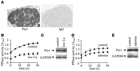
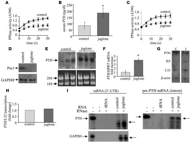
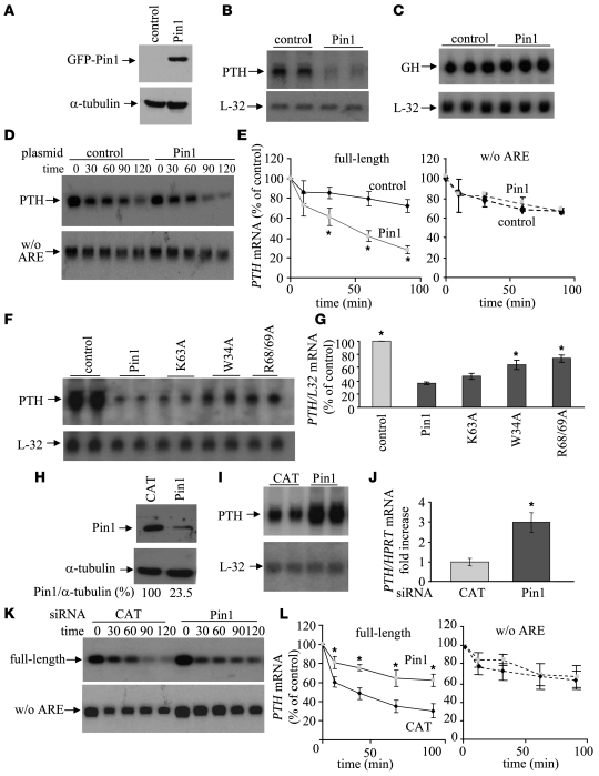
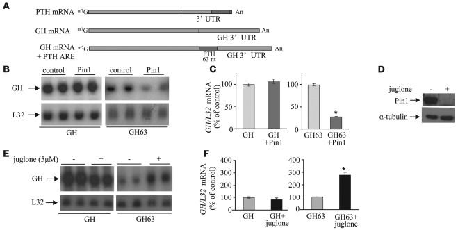
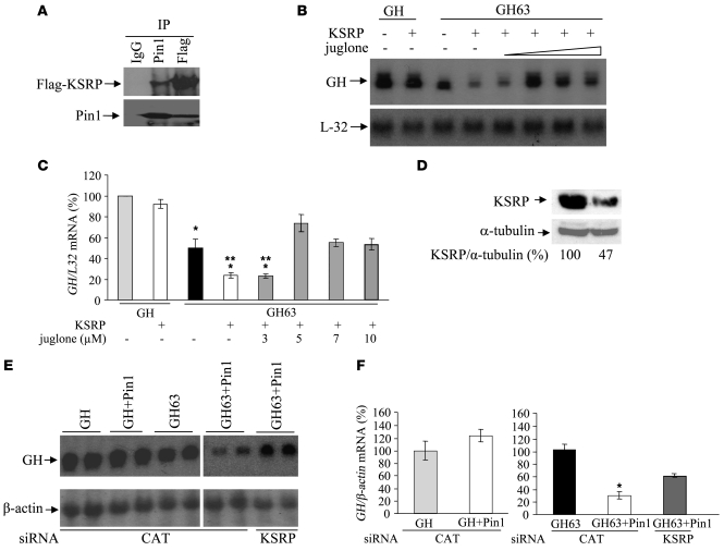
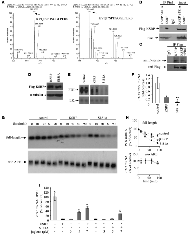
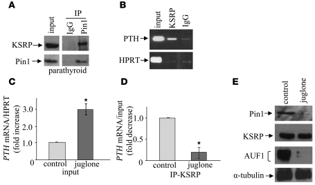
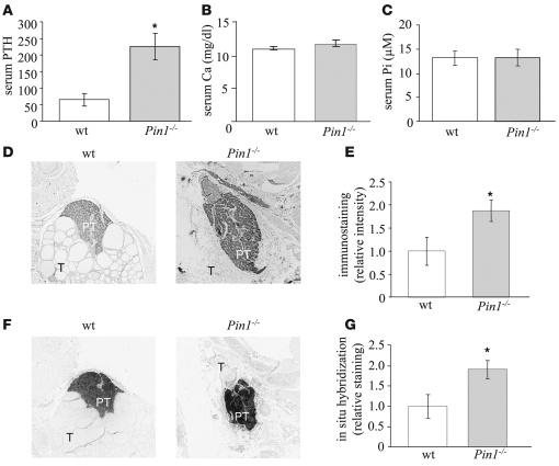
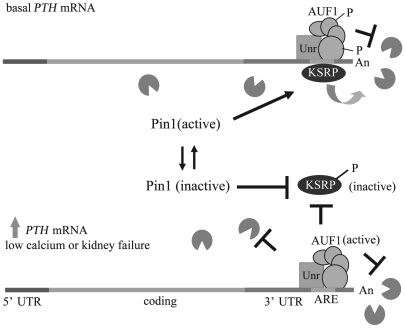
Secondary hyperparathyroidism is a major complication of chronic kidney disease (CKD). In experimental models of secondary hyperparathyroidism induced by hypocalcemia or CKD, parathyroid hormone (PTH) mRNA levels increase due to increased PTH mRNA stability. K-homology splicing regulator protein (KSRP) decreases the stability of PTH mRNA upon binding a cis-acting element in the PTH mRNA 3' UTR region. As the peptidyl-prolyl isomerase (PPIase) Pin1 has recently been shown to regulate the turnover of multiple cytokine mRNAs, we investigated the role of Pin1 in regulating PTH mRNA stability in rat parathyroids and transfected cells. The data generated were consistent with Pin1 being a PTH mRNA destabilizing protein. Initial analysis indicated that Pin1 activity was decreased in parathyroid protein extracts from both hypocalcemic and CKD rats and that pharmacologic inhibition of Pin1 increased PTH mRNA levels posttranscriptionally in rat parathyroid and in transfected cells. Pin1 mediated its effects via interaction with KSRP, which led to KSRP dephosphorylation and activation. In the rat parathyroid, Pin1 inhibition decreased KSRP-PTH mRNA interactions, increasing PTH mRNA levels. Furthermore, Pin1-/- mice displayed increased serum PTH and PTH mRNA levels, suggesting that Pin1 determines basal PTH expression in vivo. These results demonstrate that Pin1 is a key mediator of PTH mRNA stability and indicate a role for Pin1 in the pathogenesis of secondary hyperparathyroidism in individuals with CKD.


Figures

Figure 1
Figure 1. PPIase activity is decreased in parathyroid extracts from rat with secondary hyperparathyroidism due to a calcium-depleted diet or an adenine and high-phosphorus diet.
(A) Immunohistochemistry using an anti-Pin1 antibody or IgG showing specific expression of Pin1 in the parathyroid cells and not in interstitial tissue or blood vessels. Original magnification, ×200. (B and D) Pin1 isomerase assays. PPIase activity of parathyroid cytoplasmic lysates from a pool of 5 rats fed a calcium-depleted diet (B) or an adenine and high-phosphorus diet (D) compared with parathyroid extracts from control rats. The results are representative of 3 independent experiments and are presented as mean ± SEM of 3 measurements from 1 of the experiments (B) and as mean ± SEM of 3 measurements from the 3 experiments combined (D). The decrease in Pin1 activity in B and D was significant (P < 0.05) compared with the respective controls at all the time intervals studied. Control: low Ca or adenine, P < 0.05 at all time intervals. (C and E) Immunoblot analysis using anti-Pin1 or anti-GAPDH antibodies for parathyroid extracts, as in B and D.
Figure 2
Figure 2. Pin1 inhibition by juglone increases PTH mRNA in vivo posttranscriptionally.
(A and B) Juglone i.p. (A) PPIase activity assays of parathyroid cytoplasmic lysates from rats injected i.p. with juglone or vehicle. (B) Serum PTH in rats as in A. (CF) Juglone applied topically. (C) PPIase activity of parathyroid cytoplasmic lysates from rats treated with juglone or vehicle applied topically; data in A and C are presented as mean ± SEM (n = 3) from 1 of 3 repeat experiments; P < 0.05 at 5–30 seconds. (D) Immunoblots of the extracts in C for Pin1 and GAPDH. (E) Northern blots of PTH mRNA levels after juglone, in rats as in C. The lanes were run on the same gel but were noncontiguous (white line). (F) qRT-PCR analysis for PTH mRNA in rat parathyroids as in C; *P < 0.05. (G) Nuclear run-ons of PTH transcription in isolated nuclei of parathyroids from 5 rats in each group after topical juglone (J) or vehicle (C). (H) Quantification of PTH mRNA transcription in G, showing no difference in PTH transcription. The same result was obtained in a repeat experiment. (I) RNase protection analysis (RPA) of total parathyroid RNA from rats after topical juglone or vehicle. Left panels: RPA using an antisense probe for the PTH mRNA 3′ UTR or for control GAPDH mRNA. Right panel: RPA using an antisense probe for the first intron (newly transcribed pre–PTH mRNA). The intact probes before and after RNase treatment (arrows to the left and right of the gels, respectively) and hybridization with tRNA as a nonspecific control are shown. Juglone increased PTH mRNA but not the pre–PTH mRNA or GAPDH mRNA.
Figure 3
Figure 3. Pin1 regulates PTH mRNA steady-state levels and stability in transfected cells.
(AF) Pin1 overexpression. HEK293 cells were transiently cotransfected with expression plasmids for human PTH or GH and for GFP-Pin1 or control pcDNA3 plasmids. (A) Immunoblots of protein extracts at 48 hours for Pin1 and α-tubulin. (B) Northern blots for PTH mRNA. (C) Northern blots for GH mRNA. (D) IVDA for polyadenylated full-length rat PTH mRNA (top) or polyadenylated PTH mRNA without the ARE (bottom) and extracts from cells transfected with GFP-Pin1 or control plasmids. (E) Quantification of the IVDAs in D and 2 additional IVDAs; mean ± SEM of intact transcript; *P < 0.05. (F and G) Cells were transiently cotransfected with expression plasmids for PTH and GFP-Pin1 or Pin1 mutants at the WW domain (W34A) or at the PPIase domain (K63A,R68/69A). (F) Northern blots for PTH mRNA. (G) Quantification of 3 Northern blots; mean ± SEM; *P < 0.05. (HL) Pin1 knockdown. Cells were cotransfected with a human PTH plasmid and either Pin1 or control CAT siRNAs. (H) Immunoblots of Pin1 and α-tubulin after transfection. (I) Northern blots for PTH mRNA. (J) Quantification by qRT-PCR analysis for PTH mRNA after Pin1 siRNA. (K) IVDA for full-length rat PTH mRNA (top) or rat PTH mRNA without the ARE (bottom) and extracts from cells with either CAT or Pin1 siRNAs. (L) Quantification of 3 IVDAs; mean ± SEM of intact transcript at time 0; *P < 0.05.
Figure 4
Figure 4. Pin1 regulates PTH mRNA levels through the PTH mRNA ARE.
(A) Schematic representation of mRNAs for PTH, GH, and chimeric reporter GH containing the PTH mRNA ARE (GH63) used for transfections. (B and C) Pin1 overexpression. HEK293 cells were transiently cotransfected with expression plasmids for either GH or a chimeric GH63 (GH63) and GFP-Pin1 or pcDNA3 (control). (B) Northern blot analysis for PTH mRNA. (C) Quantification of 2 Northern blots as in B for GH (left) and GH63 (right); mean ± SEM; *P < 0.05 compared with GH63 without Pin1. (DF) Effect of juglone on GH63. Cells were transiently transfected with expression plasmids for either GH or GH63 and incubated with juglone or vehicle (–). (D) Immunoblots for Pin1 in cell extracts after juglone or vehicle. (E) Northern blots for GH mRNA. (F) Quantification of Northern blots of 3 independent experiments as in E (*P < 0.05). The lanes in B and E were run on the same gel but were noncontiguous (white lines).
Figure 5
Figure 5. Pin1 interacts with the PTH mRNA decay–promoting protein KSRP, and the effects of Pin1 and KSRP on the PTH mRNA ARE are interdependent.
(A) Pin1-KSRP interaction. HEK293 cells were transiently transfected with FLAG-KSRP plasmid and extracts immunoprecipitated with anti-FLAG or anti-Pin1 antibodies or IgG, run on SDS PAGE, and immunoblotted with the corresponding antibodies. (B and C) Cells were cotransfected with either GH or GH63 and KSRP or control pcDNA3 plasmids. After 48 hours, increasing concentrations of juglone were added for an additional 4 hours. (B) Northern blots for GH mRNA. (C) Quantification of 3 Northern blots as in B; *P < 0.05 compared with GH mRNA without KSRP; **P < 0.05 compared with GH63 mRNA without KSRP. (DF) KSRP knockdown. Cells were cotransfected with either GH or GH63 and GFP-Pin1 or control pcDNA3 plasmids together with either CAT or KSRP siRNAs. (D) Immunoblots for KSRP and α-tubulin protein levels 48 hours after transfection, showing decreased KSRP in KSRP siRNA–transfected cells. Densitometry analysis of KSRP corrected for α-tubulin is shown below the gels. (E) Northern blots for GH mRNAs. The lanes were run on the same gel but were noncontiguous (white line). (F) Quantification of 3 Northern blots as in E for GH (left) and GH63 (right); *P < 0.05. Data in C and F are represented as mean ± SEM.
Figure 6
Figure 6. Identification of KSRP phosphorylation at S181 and its role in KSRP-Pin1 interaction and PTH mRNA regulation.
(A) Mass spectrometry. MS spectra of tryptic mixture of immunoprecipitated FLAG-KSRP from transfected cells. The peak at 677.8555 m/z was assigned as the unphosphorylated form of amino acids 177–197 (left), and the peak at 717.8390 m/z the phosphorylated form of the peptide (right). Asterisk indicates serine phosphorylation. The P probability of the assignment was equal to 7.51E-06. Sequest X score was equal to 3.25. (B) KSRP-Pin1 interaction. HEK293 cells were cotransfected with either FLAG-KSRP or FLAG-KSRP S181A and GFP-Pin1 or control plasmid. Extracts were immunoprecipitated with either anti-Pin1 or IgG. The pellets and input were immunoblotted as indicated. (C) KSRP dephosphorylation. Cells were cotransfected with FLAG-KSRP and GFP-Pin1 or control plasmid. Extracts were immunoprecipitated with anti-FLAG or IgG and immunoblotted with anti–phosphor-serine or anti-FLAG antibody. (DH) Cells were cotransfected with the PTH plasmid or control and either FLAG-KSRP or FLAG-KSRP S181A. (D) Immunoblots showing equal amounts of KSRP. (E) Northern blots of PTH mRNA. The lanes were run on the same gel but were noncontiguous (white line). (F) qRT-PCR for PTH mRNA as in E. *Compared with control; **compared with KSRP. (G) IVDA using cell extracts and full-length human PTH mRNA (top) or PTH mRNA without the ARE (bottom). (H) Quantification of IVDA results in 3 experiments; mean ± SEM of time 0; KSRP compared with both control and S181A; *P < 0.05. (I) Cells were cotransfected with the PTH and KSRP or S181A plasmids with and without juglone. RNA was analyzed by qRT-PCR for PTH mRNA; *P < 0.05 (n = 3) compared with cells expressing KSRP and not treated with juglone.
Figure 7
Figure 7. Pin1 interacts with KSRP and regulates KSRP–PTH mRNA interaction in parathyroid glands.
(A) Pin1-KSRP interaction in the parathyroid. Pin1 was immunoprecipitated from microdissected parathyroid extracts using anti-Pin1 antibody or IgG, fractionated on SDS PAGE, and analyzed by immunoblots for KSRP and Pin1. The lanes were run on the same gel but were noncontiguous (white line). (BD) RIP analysis. Parathyroid glands were subjected to juglone or vehicle applied topically. RIP was performed on rat parathyroid glands using either anti-KSRP or control IgG. RNA from total lysate (input) and KSRP (IP-KSRP) or control IgG–bound fractions was assayed by qRT-PCR for PTH and control HPRT mRNAs. (B) Agarose gel of qRT-PCR products for PTH and HPRT. The size of PCR products was consistent with the predicted lengths of the amplified fragments. (C) PTH mRNA levels in the input. (D) mRNA levels in immunoprecipitated samples, corrected for mRNA levels in input; mean ± SEM of 3 experiments using parathyroids from pools of different rats. (E) Immunoblots of the parathyroid extracts as in C (input). *P < 0.05.
Figure 8
Figure 8. Pin–/– mice have high serum PTH and PTH mRNA levels.
(AC) Serum PTH, calcium, and phosphate levels in WT (n = 18) and Pin1–/– (n = 14) mice. (D) Immunostaining of thyroparathyroid section for PTH. (E) Quantification of data presented in 6 sections from each group. (F) In situ hybridization for PTH mRNA in thyroparathyroid sections. (G) Quantification of the in situ hybridization staining in 6 sections from each group. Original magnification, ×100 (D and F). *P < 0.05. PT, parathyroid; T, thyroid tissue.
Figure 9
Figure 9. Model for the regulation of PTH mRNA stability by PTH mRNA 3′ UTR ARE binding proteins.
Under basal conditions, there is a balanced interaction of the PTH mRNA with its stabilizing proteins AUF1 (isoforms p37, p40, p42 and p45) and Unr and the destabilizing protein KSRP. In hypocalcemia or CKD, Pin1 is inactive, resulting in KSRP phosphorylation and hence its inactivation. This would allow AUF1 and Unr to bind the PTH mRNA 3′ UTR ARE with a greater affinity, leading to increased PTH mRNA stability.

Comment in

References

    1. Silver, J., Naveh-Many, T., and Kronenberg, H.M. 2002. Parathyroid hormone: molecular biology. InPrinciples of bone biology . J.B. Bilezikian, L.G. Raisz, and G.A. Rodan, editors. Academic Press. San Diego, California, USA. 407–422.
    1. Moallem E., Silver J., Kilav R., Naveh-Many T. RNA protein binding and post-transcriptional regulation of PTH gene expression by calcium and phosphate. J. Biol. Chem. 1998;273:5253–5259. doi: 10.1074/jbc.273.9.5253. - DOI - PubMed
    1. Levi R., et al. Increased parathyroid hormone gene expression in secondary hyperparathyroidism of experimental uremia is reversed by calcimimetics: correlation with posttranslational modification of the trans acting factor AUF1. J. Am. Soc. Nephrol. 2006;17:107–112. doi: 10.1681/ASN.2005070679. - DOI - PubMed
    1. Kilav R., Silver J., Naveh-Many T. A conserved cis-acting element in the parathyroid hormone 3′-untranslated region is sufficient for regulation of RNA stability by calcium and phosphate. J. Biol.Chem. 2001;276:8727–8733. doi: 10.1074/jbc.M005471200. - DOI - PubMed
    1. Kilav R., Bell O., Le S.Y., Silver J., Naveh-Many T. The parathyroid hormone mRNA 3′-untranslated region AU-rich element is an unstructured functional element. J. Biol. Chem. 2004;279:2109–2116. doi: 10.1074/jbc.M305302200. - DOI - PubMed

Publication types

MeSH terms