. 2009 Oct;119(10):2898-913.
doi: 10.1172/JCI36498. Epub 2009 Sep 21.

CD4+CD25+Foxp3+ Tregs resolve experimental lung injury in mice and are present in humans with acute lung injury

Affiliations

CD4+CD25+Foxp3+ Tregs resolve experimental lung injury in mice and are present in humans with acute lung injury

Franco R D'Alessio et al. J Clin Invest. 2009 Oct.

Abstract

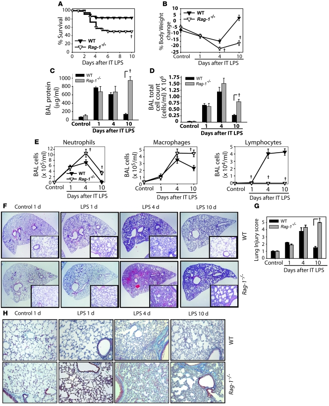
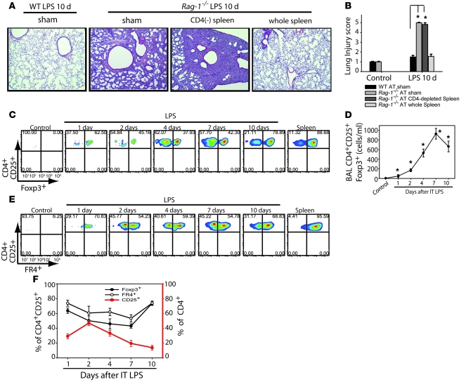
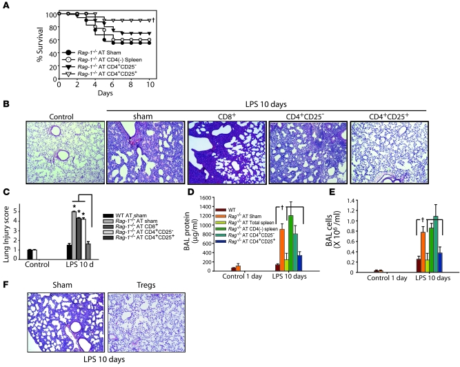
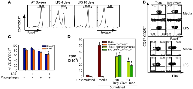
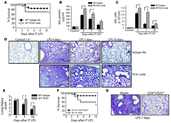
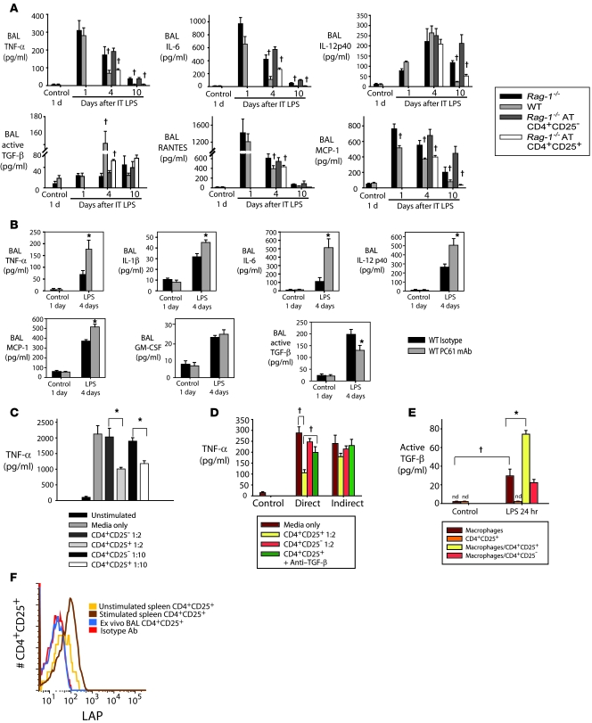
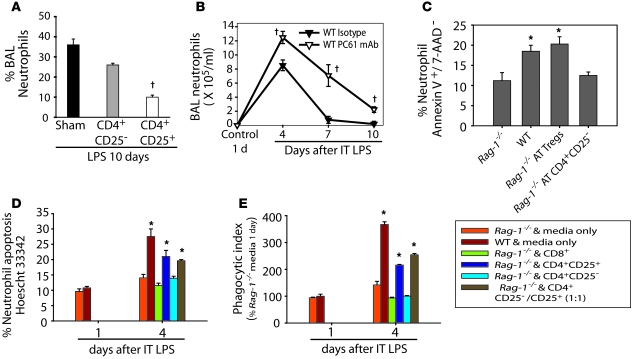
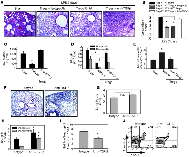
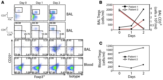
Acute lung injury (ALI) is characterized by rapid alveolar injury, inflammation, cytokine induction, and neutrophil accumulation. Although early events in the pathogenesis of ALI have been defined, the mechanisms underlying resolution are unknown. As a model of ALI, we administered intratracheal (i.t.) LPS to mice and observed peak lung injury 4 days after the challenge, with resolution by day 10. Numbers of alveolar lymphocytes increased as injury resolved. To examine the role of lymphocytes in this response, lymphocyte-deficient Rag-1-/- and C57BL/6 WT mice were exposed to i.t. LPS. The extent of injury was similar between the groups of mice through day 4, but recovery was markedly impaired in the Rag-1-/- mice. Adoptive transfer studies revealed that infusion of CD4+CD25+Foxp3+ Tregs as late as 24 hours after i.t. LPS normalized resolution in Rag-1-/- mice. Similarly, Treg depletion in WT mice delayed recovery. Treg transfer into i.t. LPS-exposed Rag-1-/- mice also corrected the elevated levels of alveolar proinflammatory cytokines and increased the diminished levels of alveolar TGF-beta and neutrophil apoptosis. Mechanistically, Treg-mediated resolution of lung injury was abrogated by TGF-beta inhibition. Moreover, BAL of patients with ALI revealed dynamic changes in CD3+CD4+CD25hiCD127loFoxp3+ cells. These results indicate that Tregs modify innate immune responses during resolution of lung injury and suggest potential targets for treating ALI, for which there are no specific therapies currently available.


Figures

Figure 1
Figure 1. Resolution of lung injury is markedly impaired in lymphocyte-deficient Rag-1–/– mice.
Mice (n = 8–10 per group per time point) were challenged with i.t. LPS. (A and B) Survival (A) and body weight relative to baseline (B) were plotted after injury. (CE) BAL total protein (C), total cell counts (D), and differential cell counts (E) were determined in WT and Rag-1–/– mice after treatment with water control or with LPS. (F) Lung sections were stained with H&E. Original magnification, ×20; ×100 (insets). (G) Histopathological mean lung injury scores from ×20 lung sections (n = 5 animals per group per time point). (H) Movat stain for collagen (blue) from WT and Rag-1–/– mice after injury. Original magnification, ×40. P < 0.05, log-rank test (mortality curves) and unpaired Student’s t test (other injury parameters).
Figure 2
Figure 2. Alveolar CD4+CD25+Foxp3+ Tregs increase after injury with i.
. LPS. (A) Representative lung sections were stained with H&E (n = 10 per group) to reveal morphologic changes on day 10 after i.t. LPS in Rag-1–/– mice. Animals received AT via tail vein injection of PBS sham treatment, 10 × 106 WT CD4-depleted splenocytes, or WT splenocytes from whole spleen. Original magnification, ×40. (B) Mean histopathological lung injury scores by day 10 after i.t. LPS (n = 6–8 animals per group). (C) BAL cells from WT animals were analyzed by flow cytometry for the presence of CD4+CD25+ surface staining and intracellular transcription factor Foxp3+ at baseline and after injury; corresponding populations in the spleen were used for comparison. (D) Absolute Treg numbers at baseline and after injury. *P < 0.05 versus control. (E) Surface staining for FR4 was determined in CD4+CD25+ cells from spleen and BAL after LPS. (F) Relative expression of Foxp3 and FR4 in CD4+CD25+ cells (left axis), as well as CD25+ expression in the CD4+ pool (right axis), isolated from the BAL at baseline and after i.t. LPS. Numbers within plots in C and E denote the percentage of cells in the respective quadrants.
Figure 3
Figure 3. AT of Tregs mediates resolution of lung injury in Rag-1–/– mice.
Rag-1–/– mice were challenged with i.t. LPS and 1 hour afterward received PBS sham treatment or 1.0 × 10⁶ WT CD4+CD25 or WT CD4+CD25+ splenocytes. (A) Rag-1–/– mouse survival over a 10-day period. P < 0.05 versus sham control, log-rank test. (B) H&E stain of representative lung sections on day 10 after i.t. LPS and infusion of PBS or the indicated lymphocyte subsets. Original magnification, ×40. (C) Mean histopathological lung injury scores (n = 8–10 animals per group). *P < 0.05. (D and E) BAL total protein (D) and total cell counts (E) were determined in WT and Rag-1–/– mice on day 10 after AT (n = 10 per group). P < 0.05 versus Rag-1–/– sham control. (F) Lung H&E staining demonstrate that AT of Tregs into injured Rag-1–/– mice as late as 24 hours after i.t. LPS achieved resolution of lung injury (n = 5 per group). Original magnification, ×40.
Figure 4
Figure 4. Phenotype and function of alveolar Tregs during resolution of ALI.
(A) Histograms showing the percentage of Foxp3+ cells in the pool of donor CD4+CD25+ splenocytes (AT spleen) as well as in CD4+CD25+ cells recovered in the BAL from LPS-injured Rag-1–/– mice on days 4 and 10 after LPS. Labeling for isotype control Ab at day 10 is also shown. (B) Purified Tregs from naive WT spleens were cultured in the presence or absence of 100 ng/ml LPS and/or macrophages. Representative flow cytometry shows the relationship between Foxp3 and FR4 expression under different conditions. Numbers within plots denote the percentage of cells in the respective quadrants. (C) Relative expression of Foxp3 and FR4 in CD4+CD25+ cells from the experiments in B. P < 0.05 versus Tregs without LPS or macrophages. (D) Proliferative responses of CD4+CD25 lymphocytes to 0.5 mg/ml anti-CD3 and irradiated APCs (Stimulated) in the presence or absence of CD4+CD25+ cells isolated from spleen or sorted from BAL of WT mice on day 4 after LPS and cultured at the indicated ratios. Unstimulated CD4+CD25 lymphocytes were not exposed to anti-CD3 and irradiated APCs. P < 0.05 versus media-stimulated CD25 cells.
Figure 5
Figure 5. Manipulation of Tregs in WT mice determines resolution of lung injury.
WT mice were pretreated with 0.5 mg of CD25-depleting PC61 mAb or isotype Ab i.p. on days –2, 0, 3, and 6 relative to i.t. LPS (day 0). (A) Survival curves over 10 days (n = 20 per group). (B and C) BAL total protein (B) and total cell counts (C) were determined at the indicated time points (n = 6 per group per time point). (D) H&E stain of representative lung sections at the indicated times. Original magnification, ×40. (E) Mean lung injury scores (n = 4 per group per time point). (F) To assess the effect of increasing Treg number, WT mice were challenged with twice our standard i.t. LPS dose (7.5 mg/kg) and 1 hour later given an infusion of 2 × 10⁶ Tregs or 100 μl PBS sham treatment, and survival was assessed (n = 10 per group). (G) Representative H&E-stained lung sections on day 7 after LPS in mice receiving PBS or Tregs. Original magnification, ×40. P < 0.05 versus respective control, log-rank test (mortality curves) and unpaired Student’s t test (other parameters).
Figure 6
Figure 6. Tregs alter alveolar cytokine profiles after LPS-induced lung injury.
WT and Rag-1–/– mice received LPS or sterile water as control i.t. (A) Rag-1–/– mice received CD4+CD25 or CD4+CD25+ cells by tail vein injection 1 hour after LPS. BAL was harvested at the indicated times and assayed for the indicated cytokines. P < 0.05 versus respective Rag-1–/– value. (B) BAL cytokines were assessed on day 4 after i.t. LPS in mice receiving isotype Ab or PC61. *P < 0.05 versus respective isotype value. (C) Primary alveolar macrophages were isolated from unstimulated WT mice and exposed to 10 ng/ml LPS. (D) Cocultures of alveolar macrophages and the indicated T cells (1:2 lymphocyte/macrophage ratio) was performed both directly (in the bottom chamber of a Transwell plate) and indirectly (macrophages and T cells in the bottom and top chambers, respectively), and TNF-α was measured 24 hours after LPS stimulation. (E) Thioglycollate-induced peritoneal macrophages were harvested, plated on 24-well plates (1 × 10⁶ cells/well), and cocultured with CD4+CD25+ or CD4+CD25 cells or with media alone at a 1:2 lymphocyte/macrophage ratio in the presence or absence of 10 ng/ml LPS. Active TGF-β was measured in supernatants by ELISA. (CE) P < 0.05; *P < 0.05. (F) Cell surface expression of LAP in CD4+CD25+ splenocytes incubated with medium (unstimulated) or cultured with 10 μg/ml anti-CD3 and 50 U/ml IL-2 (stimulated), ex vivo BAL CD4+CD25+ cells on day 4 after i.t. LPS, and isotype Ab control.
Figure 7
Figure 7. Tregs facilitate alveolar neutrophil clearance in vivo and in vitro.
(A) After administration of i.t. LPS and AT of PBS sham treatment or the indicated lymphocyte subtypes, the percentage of neutrophils in the BAL on day 10 was assessed. P < 0.05 vs. sham. (B) Percent neutrophils in the alveolar compartment of WT mice after receiving isotype or Treg-depleting Abs. P < 0.05 versus isotype Ab. (C) Percent BAL neutrophils undergoing apoptosis, as assessed by dual labeling with Annexin V and 7-AAD on day 4 after i.t. LPS in WT or Rag-1–/– mice receiving PBS or AT of Tregs or CD4+CD25 cells. (D) WT and Rag-1–/– mice were exposed to i.t. LPS, and on day 1 or day 4, both BAL and ex vivo BAL mixed cultures were performed. Neutrophils and macrophages were cultured in vitro for 6 hours, then media or the indicated lymphocyte subset was added in a 1:10 lymphocyte/macrophage ratio. (E) Neutrophil apoptosis or macrophage phagocytosis was assessed 24 hours after addition of the indicated cells in vitro. *P < 0.05 vs. day-1 Rag-1–/– and media only.
Figure 8
Figure 8. TGF-β blockade abrogates Treg-mediated effects on resolution of lung injury.
PBS sham treatment or AT of 1 x 10⁶ Tregs (isolated from WT or IL-10–/– mice) was performed via tail vein injection (day 0) into i.t. LPS–injured Rag-1–/– mice. Anti–TGF-β Ab (150 μg/dose per mouse on days 0 and 4) or isotype Ab were administered. (A) H&E stain of representative lung sections on day 7 after i.t. LPS (n = 5 per group). Original magnification, ×40. (B) Mean histopathological lung injury scores (n = 4 per group). (CE) BAL total protein (C) and total cell counts (D) were determined for groups as designated. BAL neutrophil apoptosis (E) was measured by flow cytometry using dual labeling with Annexin V/ 7-AAD. n = 10 per group. (F) Anti–TGF-β or isotype Abs were administered to WT mice on days 0, 2, and 4 after i.t. LPS, and lung histology was examined on day 7. Original magnification, ×20. (G) Mean histopathological lung injury scores for samples from F (n = 4 per group). P = 0.1. (H) BAL total cell and neutrophil counts. (I and J) After gating on Ly6G cells, decreased BAL neutrophil apoptosis was observed by staining with Annexin V and 7-AAD. (J) Flow cytometry of Ly6G+ cells (neutrophils) labeled with Annexin V and 7-AAD. Numbers within plots denote the percentage of cells in the respective quadrants. *P < 0.05 versus respective sham control; P < 0.05 versus respective isotype control.
Figure 9
Figure 9. Alveolar Tregs are present in humans with ALI.
Mini-BAL and blood sampling was performed on days 0, 1, and 2 relative to enrollment. (A) Patient 1 BAL flow cytometry panels for CD3+, CD4+ and CD8+, and CD3+CD4+CD25hiCD127loFoxp3+ cells, as well as flow cytometry for CD3+CD4+CD25hiCD127loFoxp3+ cells in the blood compartment. Intracellular staining with human isotype control Ab for FoxP3, in cells gated as described above, is also shown. Numbers within plots denote the percentage of cells in the respective quadrants or gated regions. (B) Absolute number of CD3+CD4+CD25hiCD127loFoxp3+ Tregs in the BAL (right axis) and blood (left axis) over time in patients with ALI. (C) Absolute number of CD3+CD4+CD25hiCD127loFoxp3+ Tregs in the blood of patients with ALI.

Comment in

References

    1. Ware L.B., Matthay M.A. The acute respiratory distress syndrome. N. Engl. J. Med. 2000;342:1334–1349. doi: 10.1056/NEJM200005043421806. - DOI - PubMed
    1. Rubenfeld G.D., et al. Incidence and outcomes of acute lung injury. N. Engl. J. Med. 2005;353:1685–1693. doi: 10.1056/NEJMoa050333. - DOI - PubMed
    1. Serhan C.N., Savill J. Resolution of inflammation: the beginning programs the end. Nat. Immunol. 2005;6:1191–1197. doi: 10.1038/ni1276. - DOI - PubMed
    1. Savill J. Apoptosis in resolution of inflammation. J. Leukoc. Biol. 1997;61:375–380. - PubMed
    1. Henson P.M. Dampening inflammation. Nat. Immunol. 2005;6:1179–1181. doi: 10.1038/ni1205-1179. - DOI - PubMed

Publication types

MeSH terms