. 2009 Sep 22;4(9):e7120.
doi: 10.1371/journal.pone.0007120.

Estrogen/estrogen receptor alpha signaling in mouse posterofrontal cranial suture fusion

Affiliations

Estrogen/estrogen receptor alpha signaling in mouse posterofrontal cranial suture fusion

Aaron W James et al. PLoS One. .

Abstract

Background: While premature suture fusion, or craniosynostosis, is a relatively common condition, the cause is often unknown. Estrogens are associated with growth plate fusion of endochondral bones. In the following study, we explore the previously unknown significance of estrogen/estrogen receptor signaling in cranial suture biology.

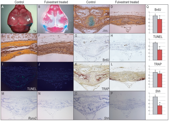
Methodology/principal findings: Firstly, estrogen receptor (ER) expression was examined in physiologically fusing (posterofrontal) and patent (sagittal) mouse cranial sutures by quantitative RT-PCR. Next, the cranial suture phenotype of ER alpha and ER beta knockout (alphaERKO, betaERKO) mice was studied. Subsequently, mouse suture-derived mesenchymal cells (SMCs) were isolated; the effects of 17-beta estradiol or the estrogen antagonist Fulvestrant on gene expression, osteogenic and chondrogenic differentiation were examined in vitro. Finally, in vivo experiments were performed in which Fulvestrant was administered subcutaneously to the mouse calvaria. Results showed that increased ERalpha but not ERbeta transcript abundance temporally coincided with posterofrontal suture fusion. The alphaERKO but not betaERKO mouse exhibited delayed posterofrontal suture fusion. In vitro, addition of 17-beta estradiol enhanced both osteogenic and chondrogenic differentiation in suture-derived mesenchymal cells, effects reversible by Fulvestrant. Finally, in vivo application of Fulvestrant significantly diminished calvarial osteogenesis, inhibiting suture fusion.

Conclusions/significance: Estrogen signaling through ERalpha but not ERbeta is associated with and necessary for normal mouse posterofrontal suture fusion. In vitro studies suggest that estrogens may play a role in osteoblast and/or chondrocyte differentiation within the cranial suture complex.


Conflict of interest statement

Competing Interests: The authors have declared that no competing interests exist.

Figures

Figure 1
Figure 1. Estrogen receptor α and β expression in PF and SAG sutures.
(A) ERα gene expression in PF and SAG sutures by qRT-PCR, normalized to GAPDH. Expression increased over 12-fold from postnatal ages 4 to 10 in the fusing PF suture, see red bars. Highest expression was noted at p10, temporally corresponding with PF suture fusion. (B) ERβ gene expression in PF and SAG sutures, normalized to GAPDH. Expression remained relatively constant over the time course examined in both PF and SAG sutures. (C) Sox9 gene expression in PF and SAG sutures, normalized to GAPDH. (D) Pentachrome staining of the p7 PF suture in which cartilage appears blue/green, while bone appears yellow. (E) ERα immunohistochemistry of p7 PF suture. Nuclear ERα protein strongly localizes to hypertrophic chondrocytes within the PF suture; dashed lines indicate the fusing inner bone table of the suture. (F) Negative control for ERα immunohistochemistry. (G) Pentachrome staining of the p7 SAG suture. (H) ERα immunohistochemistry of p7 SAG suture. Dashed lines indicate the adjacent osteogenic fronts of the patent suture. (I) Negative control for ERα immunohistochemistry. Gene expression values are normalized to expression within the p4 SAG suture complex, N = 10, *, **, and #P≤0.01 in which * signifies differences between the PF suture at various timepoints, ** between SAG suture at various timepoints, and # between PF and SAG sutures at the same timepoint. Histological sections are from the anterior aspect of PF and SAG sutures, presented at 40× magnification.
Figure 2
Figure 2. PF suture phenotype in αERKO mouse.
(A) Wild-type (WT) p10 PF suture. Hypertrophic chondrocytes, seen here as blue/green, are observed to bridge the gap between osteogenic fronts of the suture. (B) αERKO p10 PF suture. A complete absence of cartilage is observed, despite proximity of the osteogenic fronts. (C) PCNA immunohistochemistry in the WT p10 PF suture. (D) PCNA staining in the αERKO p10 PF suture showed relatively less staining. (E) In situ hybridization for Runx2 in the WT p10 frontal bone. Strong staining was noted in the cells lining and within the bone. (F) In comparison, less staining was observed in the αERKO frontal bone (G) In situ hybridization for Shh in the WT p10 PF suture. Strong staining was noted in the hypertrophic chondrocytes within the suture mesenchyme. (H) In comparison, little staining for Shh was noted in the αERKO p10 PF suture. (I) WT p16 PF suture. Note normal osseous fusion on the endocranial aspect. (J) αERKO p16 PF suture. Suture patency is apparent on both endo- and ectocranial aspects. (K) Mean percentage suture fusion in WT, αERKO and βERKO PF p16 sutures. Every 5th coronal section of the suture was stained; two blinded, independent observers judged fusion or patency along the length of the suture. (L) Relative gene expression in WT as compared to αERKO whole skulls at 7d of life, as determined by qRT-PCR. Averages and standard deviations were calculated, significance calculated relative to wild-type percentage fusion, *P<0.01. For histological specimens, sections presented are taken from the anterior aspect of the PF suture, presented at 20×–40× magnification.
Figure 3
Figure 3. Cellular proliferation of PF SMCs with 17-β Estradiol and Fulvestrant.
After 3 and 6d growth with or without 17-β estradiol (0.1–100 nM), BrdU incorporation assays were performed to evaluate cellular proliferation. (A) BrdU incorporation with or without 17-β estradiol. Treatment with 17-β estradiol (E2) resulted in increased proliferation (0.1–10 nM). (B) BrdU incorporation after 6d growth with Fulvestrant alone or in combination with 17-β estradiol. Fulvestrant alone showed no effect on BrdU uptake (left), while in combination with E2 was observed to reverse the mitogenic effect of E2 (right). Values are normalized and significance levels calculated relative to control groups in grey, N = 6, *P<0.01.
Figure 4
Figure 4. Osteogenic differentiation of PF SMCs with 17-β Estradiol.
(A) ERα and ERβ expression throughout osteogenic differentiation (0, 1 and 2 wks). (B, left) Enzymatic alkaline phosphatase activity normalized to total protein content after 7d differentiation with or without E2. (B, right) Photometric quantification of Alizarin red staining after 14d differentiation with or without E2. (C) Alkaline phosphatase staining with or without E2 after 7d differentiation. (D) Alizarin red staining with or without E2 after 14d differentiation. (E, left) Expression of early markers of osteogenic differentiation (Runx2, ALP and Col1α) at 4d differentiation with or without E2. (E, right) Osteocalcin expression after 14d differentiation with or without E2. (F) Bmp2, Bmp4 and Bmp7 expression at 4d differentiation with or without E2. (G) Shh, Ihh, Gli1 and Ptc1 expression at 4d differentiation with or without E2. Values are normalized and significance levels calculated relative to control groups in grey or 0 wk expression levels in blue, photos are representative of random microscopical fields at 20× magnification, N = 3, *P<0.01.
Figure 5
Figure 5. Osteogenic differentiation of PF SMCs with Fulvestrant.
(A) Alkaline phosphatase staining with or without Fulvestrant after 7d differentiation. (B) Alizarin red staining with or without Fulvestrant after 14d differentiation. (C) Enzymatic alkaline phosphatase activity normalized to total protein content after 7d differentiation with or without Fulvestrant. (D, left) Runx2 and Col1a expression after 4d differentiation with or without Fulvestrant. (D, right) Osteopontin and Osteocalcin expression after 14d differentiation with or without Fulvestrant. (E) Bmp2, Bmp4, and Bmp7 expression after 4d with or without Fulvestrant. (F) Shh, Ihh, Gli1, and Ptc1 after 4d differentiation with or without Fulvestrant. Values are normalized and significance levels calculated relative to control groups in grey, photos are representative of random microscopical fields at 20× magnification, N = 3, *P<0.01.
Figure 6
Figure 6. Chondrogenic differentiation of PF SMCs with 17-β Estradiol.
(A–B) ERα and ERβ mRNA expression overtime during in vitro chondrogenic differentiation. Both transcripts were observed to increased overtime in culture after 6d differentiation. (C) Sox9 expression during chondrogenic differentiation with or without E2 (10 nM). (D) Col II expression with or without E2 (10 nM). (E) Tgf-β1 expression with or without E2 (10 nm). Values are normalized and significance levels calculated relative to control groups or 0wk expression in grey or blue, N = 3, *P<0.01.
Figure 7
Figure 7. Fulvestrant application to mouse calvaria.
(A–B) Control treated and Fulvestrant treated calvariae (N = 6, N = 10 respectively) (A) Dorsal view of p10 bone and cartilage preparation, showing normal calvarial morphogenesis in vehicle treated calvaria. (B) Overhead view of comparable Fulvestrant treated calvaria. A significant delay in mineralization was observed with widened sutures. (C) Pentachrome staining of p10 PF suture sectioned in a coronal plane. Normal developmental timing is observed, including progressive fusion and presence of a cartilage intermediate. (D) Comparable histological section of Fulvestrant treated a p10 PF suture, patency and diminished osteoid is apparent without cartilage. (E–F) Image of mid-frontal bone in control as compared to Fulvestrant treated calvaria, pentachrome stain. Severely diminished osteoid deposition (appearing yellow) is apparent among Fulvestrant treated calvaria. (G–H) BrdU incorporation in control in comparison to treated PF sutures. BrdU positive cells appear brown, while nuclei are counterstained with haemotoxylin. No significant difference was observed between control and Fulvestrant treated sutures (I–J) TUNEL staining in control and Fulvestrant treated PF sutures. Positive cells appear green. (K–L) TRAP staining in control and Fulvestrant treated PF sutures. Positively staining cells appear red. (M–N) Runx2 in situ hybridization in control and Fulvestrant treated PF sutures. Positively staining cells appear purple. (O–P) Shh in situ hybridization in control and Fulvestrant treated PF sutures. Positively staining cells appear purple. (Q) Quantitative assessments of percentage +BrdU, +TUNEL, or +TRAP stained cells, as calculated using Adobe Photoshop. For quantitative assessments, values are normalized and significance levels calculated relative to control treated groups shown in grey, *P<0.01.

References

    1. Farkas LG, Tompson B, Phillips JH, Katic MJ, Cornfoot ML. Comparison of anthropometric and cephalometric measurements of the adult face. J Craniofac Surg. 1999;10:18–25; discussion 26. - PubMed
    1. Panchal J, Uttchin V. Management of craniosynostosis. Plast Reconstr Surg. 2003;111:2032–2048; quiz 2049. - PubMed
    1. Lenton KA, Nacamuli RP, Wan DC, Helms JA, Longaker MT. Cranial suture biology. Curr Top Dev Biol. 2005;66:287–328. - PubMed
    1. Sahar DE, Longaker MT, Quarto N. Sox9 neural crest determinant gene controls patterning and closure of the posterior frontal cranial suture. Dev Biol. 2005;280:344–361. - PubMed
    1. Manzanares MC, Goret-Nicaise M, Dhem A. Metopic sutural closure in the human skull. J Anat. 1988;161:203–215. - PMC - PubMed

Publication types