

A morphological comparison of the human lumbar multifidus by chemical dissection
- PMID: 19771186
- PMCID: PMC2716159
- DOI: 10.1179/jmt.2008.16.4.84E
A morphological comparison of the human lumbar multifidus by chemical dissection
Abstract
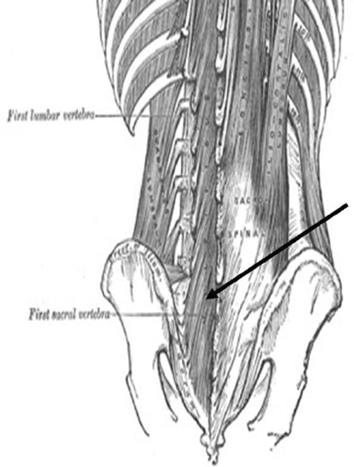
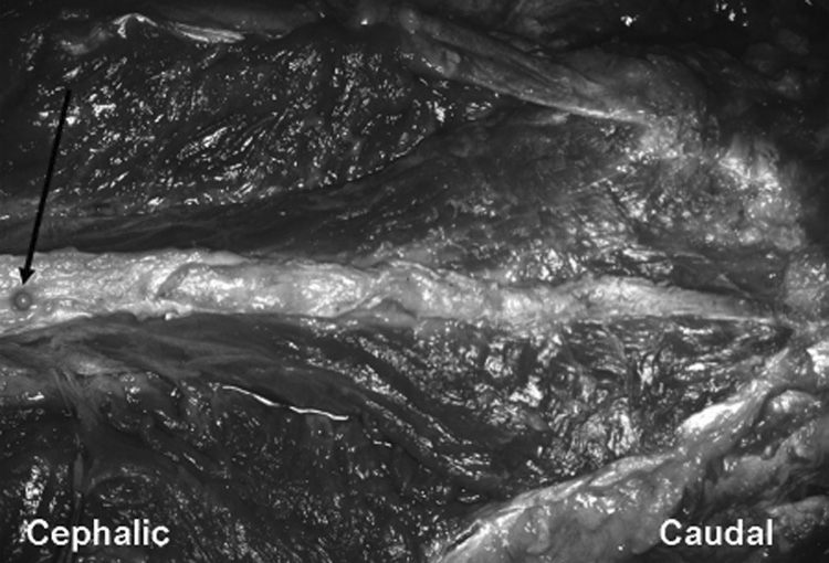
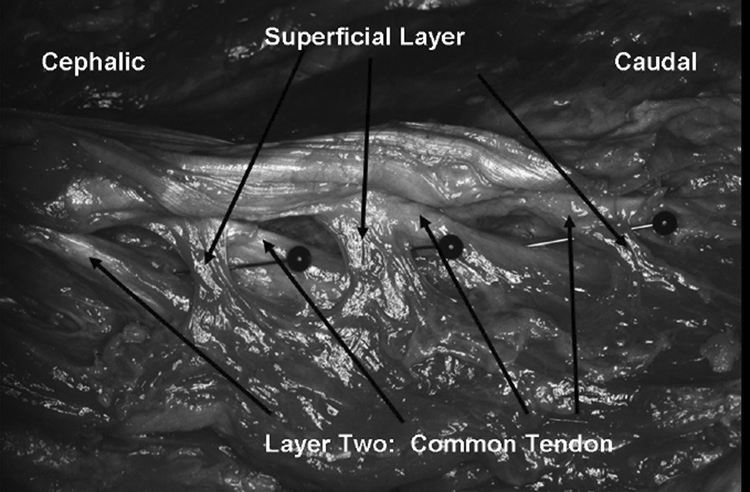
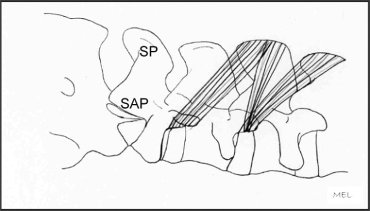
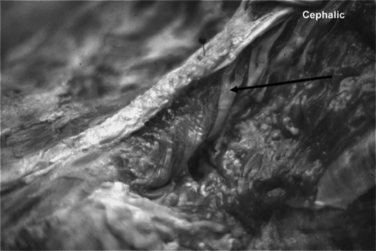
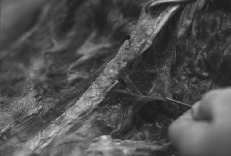
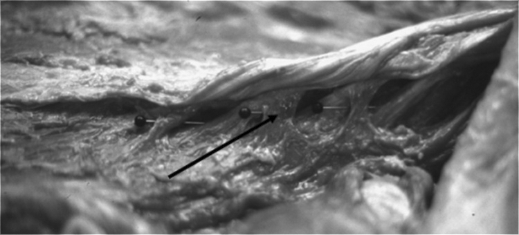
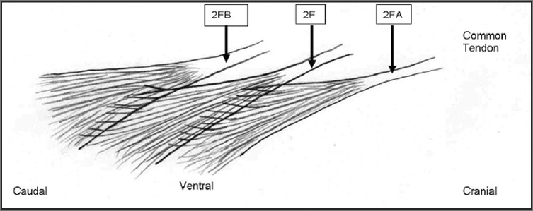
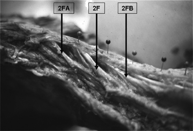
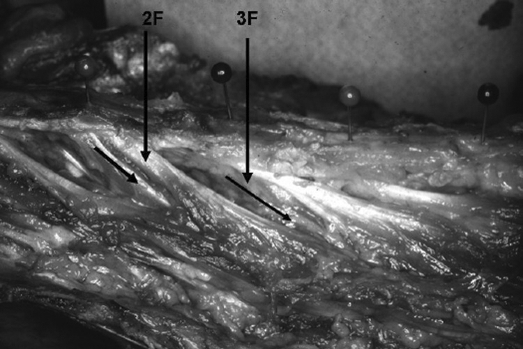
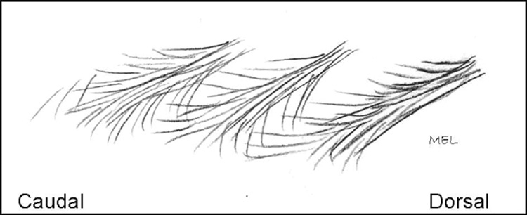
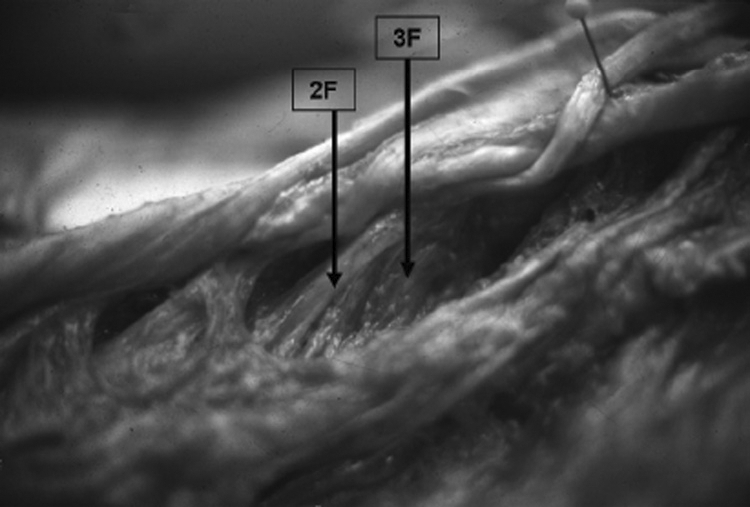
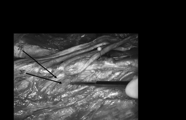
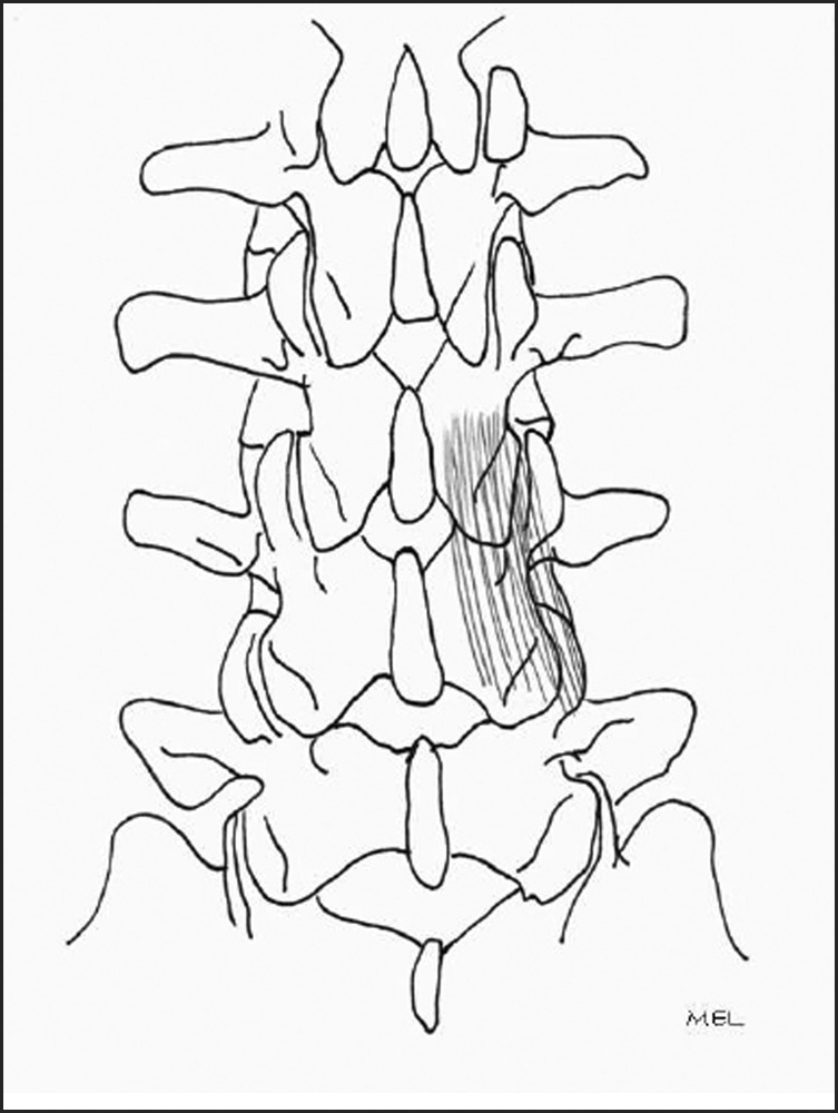
This anatomical study describes the morphology of the human lumbar multifidus muscle through gross and chemical dissection of fresh cadavers. Previous morphological descriptions were analyzed with regard to fascicular divisions and cleavage planes. Gross dissection was performed on the lumbar multifidus of four fresh adult human cadavers and four preserved cadavers. Gradual chemical dissection using nitric or formic acid was used for connective tissue digestion to enhance the documentation of muscle fiber direction. Results revealed four distinct layers of the lumbar multifidus separated by cleavage planes. The superficial layer was more extensive than previously described with bony attachments at both the origin and insertion at several vertebral levels. The attachments of the second through fourth layers differed in that distinct cleavage planes between the various fascicles were not found with chemical dissection. The lumbar multifidus has a multipennate fiber arrangement, and the fascicles between the various layers inter-attach. Inter-fascicle attachment differs with the description by Macintosh et al of distinct cleavage planes between and within the fascicles of each layer. Accurate anatomical knowledge of the fascicles of the lumbar multifidus is integral for defining the actions of this complex lumbar muscle. This study supports the clinical belief that the multifidus has a significant role in control and stabilization of the lumbar spine in multiple planes of action. The multipennate arrangement of this muscle with fascicular inter-attachment supports the clinical premise that the multifidus is activated in a variety of positions and can potentially produce and mediate intersegmental mobility and provide proprioception.
Keywords: Chemical Dissection; Lumbar Region; Multifidus; Muscles.
Figures
References
-
- Hides JA, Richardson CA, Jull GA. Multifidus muscle recovery is not automatic after resolution of acute, first-episode low back pain. Spine. 1996;21:2763–2769. - PubMed
-
- Mattila M, Hurme M, Alaranta H, et al. The multifidus muscle in patients with lumbar disc herniation: A histochemical and morphometric analysis of intraoperative biopsies. Spine. 1986;11:732–738. - PubMed
-
- Gray H. Gray's Anatomy, 36th ed. London, UK: Warwick and Williams; 1980.
-
- Cunningham DJ. Textbook of Anatomy, 12th ed. London, UK: Oxford University Press; 1981.
LinkOut - more resources
Full Text Sources