

Review
doi: 10.1021/cr9002787.
Microbial iron acquisition: marine and terrestrial siderophores
Affiliations
- PMID: 19772347
- PMCID: PMC2761978
- DOI: 10.1021/cr9002787
Item in Clipboard
Review
Microbial iron acquisition: marine and terrestrial siderophores
Chem Rev.
2009 Oct.
No abstract available
Figures

Microbial (Gram negative) iron uptake pathways.

Ribbon diagrams of outer membrane siderophore receptor proteins from E. coli: ferric-citrate (FecA), ferric-enterobactin (FepA) and ferric-hydroxamate (FhuA); and P. aeruginosa: ferric pyoverdine (FpvA) and ferric pyochelin (FptA).

Schematic of the proteins involved in ferrichrome transport. The crystal structure of FhuA in complex with the C-terminus of TonB was reported by Pawelek et al, 2006.

Ribbon representation of the S. marcescens hemophore, HasA (red), bound to its outer membrane receptor protein HasR (blue) (PDB code 3CSN).

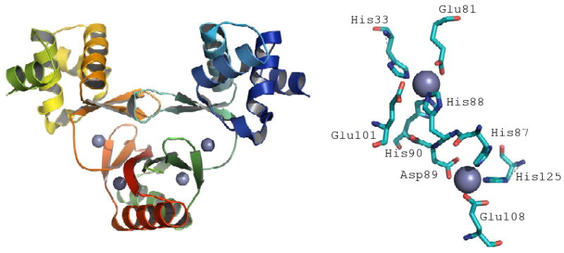
Ribbon diagram depiction of the Haemophilus influenza Fbp protein, the ferric binding site is shown on the right. The ferric ion is coordinated by two oxygens from Tyr195 and Tyr196, an imidazole nitrogen from His9, a carboxylate oxygen from Glu57, an oxygen atom from an exogeneous phosphate anion, and an oxygen atom from a water molecule in an octahedral arrangement. (PDB Code 1MRB)
Crystal structure of the ferric uptake regulator (Fur) protein from Vibrio cholerae.
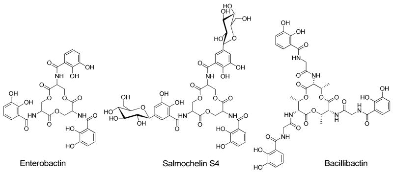
Structures of enterobactin, salmochelin S4 and bacillibactin.

Structures of desferrioxamines E, G and B.

Structures of selected α-hydroxycarboxylate siderophores.

Suites of marine amphiphilic siderophores: marinobactins (Marinobacter sp. DS40M6)- and aquachelins (Halomonas aquamarina DS40M3); amphibactins (Vibrio sp. R10); loihichelins (Halomonas sp. LOB-5); ochrobactins (Ochrobactrum sp. SP18); synechobactins (Synechococcus sp. PCC 7002).

Coordination of Fe(III) could give ME a larger head group area : tail volume ratio such that a smaller micelle is formed. Reproduced from reference 107.
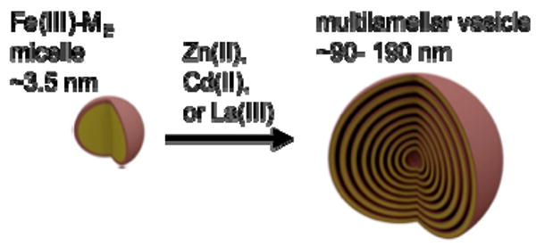
Multilamellar vesicle formation from Fe(III)-marinobactin E induced by addition of Zn(II), Cd(II), La(III) or excess Fe(III). Adapted from reference.
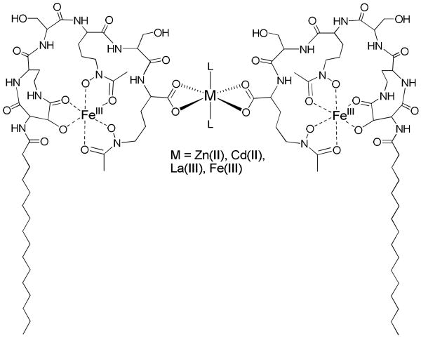
Proposed terminal carboxylate crosslinking of marinobactin E by the added cations, M (Zn(II), Cd(II), La(III) or excess Fe(III)). The bis-bidentate coordination geometry of the two carboxylates shown in the figure could also be bis-monodentate carboxylate cross linking. The resulting “composite surfactant” would have a lower headgroup-area : tail-volume ratio that may favor vesicle formation. “L” is an undefined ligand to fill out the octahedral coordination.

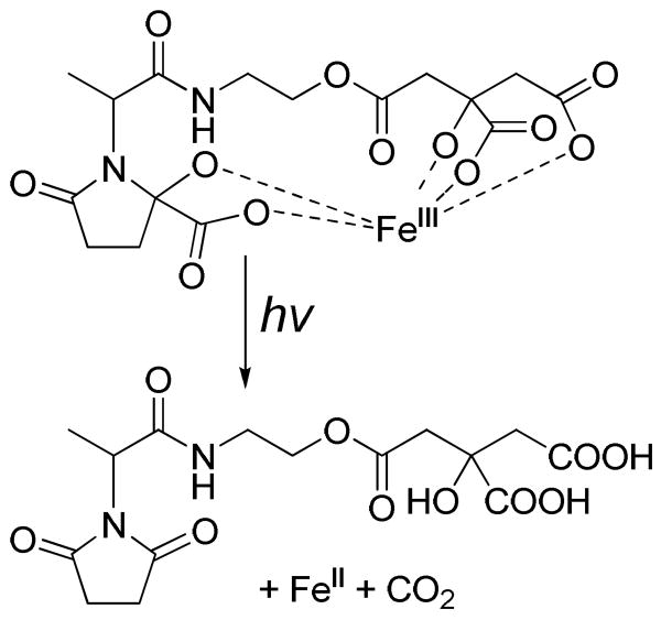
Reaction scheme for the uv photolysis of Fe(III)-aerobactin under aerobic conditions.
Photoreaction of Fe(III)-vibrioferrin. Reaction derived from data presented in reference 120.

Proposed photoreaction of diferric dicitrate in acid. Reaction derived from data presented in reference 114.

Photoreaction of Fe(III)-aquachelin. “L” is an undefined ligand to fill out the octahedral coordination.

Structures of other marine peptide siderophores that contain β-hydroxyaspartic acid.

Other siderophores produced by marine pathogens and oceanic bacteria: petrobactin, petrobactin-(SO3H), and petrobactin-(SO3H)2 (M hydrocarbonoclasticus, Marinobacter aquaeolei VT8);,, vanchrobactin and anguibactin (Vibrio anguillarum);, amonabactins (Aeromonas hydrophila).

Structures of the ornibactins and corrugatin.
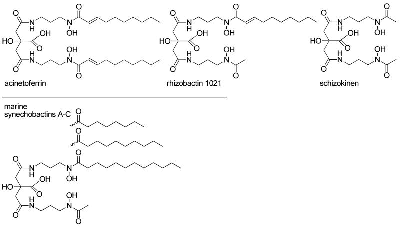
Comparison of the amphiphilic citrate siderophores of acinetoferrin rhizobactin 1021 and the synechobactins to the hydrophilic schizokinen siderophore.

Structures of mycobactins and carboxymycobactins produced by Mycobacteria.

Multiple siderophores produced by different pathogenic bacteria: enterobactin,, salmochelins, aerobactin,, and yersinabactin (E. coli, Salmonella, and Yersinia sp.); bacillibactin and petrobactin (Bacillus sp.); pyochelin and pyoverdin (P. aeruginosa); chrysobactin and achromobactin (E. chrysanthemi).,

Ribbon representation of siderocalin bound to ferric enterobactin (PDB code 3BYO).
References
-
- Crosa JH, Mey AR, Payne SM, editors. Iron Transport In Bacteria. ASM Press; Washington DC: 2004.
-
- Templeton DM, editor. Molecular and Cellular Iron Transport. Marcel Dekker, Inc.; New York: 2002.
-
- Martin JH, Gorden RM. Deep Sea Research. 1988;35:117.
-
- Aguilar-Islas AM, Hurst MP, Buck KN, Sohst B, Smith GJ, Lohan MC, Bruland KW. Progress in Oceanography. 2007;73:99.
-
- Rue EL, Bruland KW. Mar Chem. 1995;50:117.
Publication types
MeSH terms
Substances
Grants and funding
LinkOut - more resources
Full Text Sources
Other Literature Sources
Medical
