. 2009 Oct 20;48(41):9705-7.
doi: 10.1021/bi901430h.

Branch-specific sialylation of IgG-Fc glycans by ST6Gal-I

Affiliations

Branch-specific sialylation of IgG-Fc glycans by ST6Gal-I

Adam W Barb et al. Biochemistry. .

Abstract

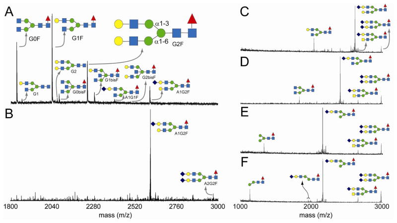
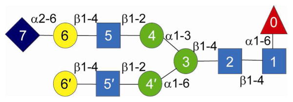
Sialylated forms of the Fc fragment of immunoglobulin G, produced by the human alpha2-6 sialyltransferase ST6Gal-I, were identified as potent anti-inflammatory mediators in a mouse model of rheumatoid arthritis and are potentially the active components in intravenous IgG anti-inflammatory therapies. The activities and specificities of hST6Gal-I are, however, poorly characterized. Here MS and NMR methodology demonstrates glycan modification occurs in a branch-specific manner with the alpha1-3Man branch of the complex, biantennary Fc glycan preferentially sialylated. Interestingly, this substrate preference is preserved when using a released glycan, suggesting that the apparent occlusion of glycan termini in Fc crystal structures does not dominate specificity.


Figures

Figure 1
Figure 1
Mass spectrometry based determination of glycan structures. (Panel A) N-glycans isolated from the immunoglobulin G Fc fragment with the G2F glycoform illustrating the α1–3Man and α1–6Man branches of the glycan. (Panel B) Galactosylation followed by sialylation of the Fc fragment results in primarily digalactosylated, monosialylated glycan. (Panels C–F) Enzymatic determination of the branch containing an N-acetylneuraminic acid residue: (Panel C) monosialylated glycan treated with β-galactosidase (Panel D) and N-acetylglucosaminidase (Panel E) resulted in the removal of those residues on the non-reducing end not protected by a terminal sialic acid. (Panel F) The sialylated, β-galactosidase- and N-acetylglucosaminidase-treated glycan was not affected by an α1–2,3 mannosidase, suggesting the terminal mannose residue is α1–6 linked. Residues are denoted by symbols: N-acetylneuraminic acid (purple diamond), galactose (yellow circle), N-acetylgalactosamine (yellow square), N-acetylglucosamine (blue square), mannose (green circle) and fucose (red triangle).
Figure 2
Figure 2
The proposed structure of the monosialylated glycan as determined by enzymatic digests and NMR spectroscopy is shown with the linkages indicated. The residue symbols are the same as those in Figure 1.

Similar articles

Cited by

References

    1. Gabius HJ. Crit Rev Immunol. 2006;26:43–79. - PubMed
    1. Kaneko Y, Nimmerjahn F, Ravetch JV. Science. 2006;313:670–673. - PubMed
    1. Anthony RM, Nimmerjahn F, Ashline DJ, Reinhold VN, Paulson JC, Ravetch JV. Science. 2008;320:373–376. - PMC - PubMed
    1. Dwyer JM. New Eng J of Med. 1992;326:107–116. - PubMed
    1. Arnold JN, Wormald MR, Sim RB, Rudd PM, Dwek RA. Anl Rev Imm. 2007;25:21–50. - PubMed

Publication types

MeSH terms