Review
. 2009 Sep 25;423(2):157-68.
doi: 10.1042/BJ20090942.

Mechanisms of double-strand break repair in somatic mammalian cells

Affiliations
Review

Mechanisms of double-strand break repair in somatic mammalian cells

Andrea J Hartlerode et al. Biochem J. .

Erratum in

  • Biochem J. 2010 Mar 15;426(3):389

Abstract

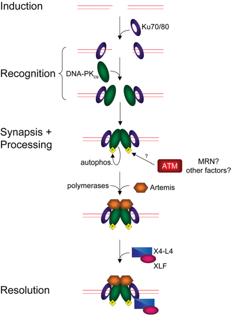
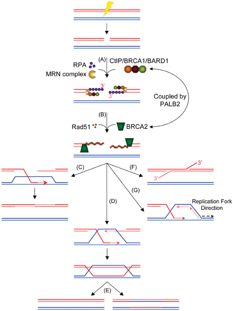
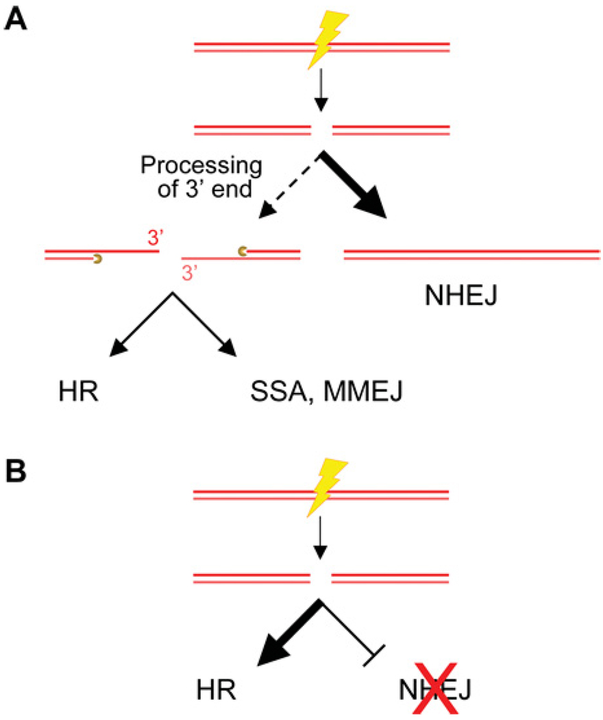
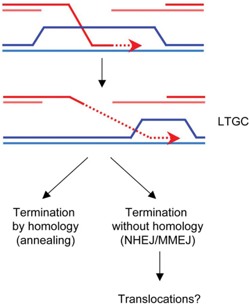
DNA chromosomal DSBs (double-strand breaks) are potentially hazardous DNA lesions, and their accurate repair is essential for the successful maintenance and propagation of genetic information. Two major pathways have evolved to repair DSBs: HR (homologous recombination) and NHEJ (non-homologous end-joining). Depending on the context in which the break is encountered, HR and NHEJ may either compete or co-operate to fix DSBs in eukaryotic cells. Defects in either pathway are strongly associated with human disease, including immunodeficiency and cancer predisposition. Here we review the current knowledge of how NHEJ and HR are controlled in somatic mammalian cells, and discuss the role of the chromatin context in regulating each pathway. We also review evidence for both co-operation and competition between the two pathways.


Figures

Figure 1
Figure 1. NHEJ in mammalian cells
Induction of a DSB forms DNA ends that are bound by the Ku heterodimer. Ku translocates inwards, allowing recruitment of DNA-PKcs to the DNA termini. The two DNA-PKcs molecules can then interact to tether the DSB ends together. Synapsis of DNA-PKcs triggers phosphorylation of DNA-PKcs [including autophosphorylation (autophos.)], altering the conformation and dynamics of DNA-PKcs. Phosphorylation of DNA-PKcs allows for recruitment of Artemis and other end-processing factors such as Pol X (DNA polymerare X) family members to generate the proper DNA ends required for ligation. Once the ends are processed, the X4-L4 complex, along with XLF, ligates the ends, repairing the break.
Figure 2
Figure 2. Homology-directed repair in eukaryotic cells
(A) Induction of a DSB is recognized by the MRN complex, which tethers the DNA ends together and participates in end processing. The CtIP–BRCA1–BARD1 complex co-operates with the MRN complex to aid in end resection. ssDNA is initially bound by the ssDNA-binding protein RPA to keep the ssDNA from forming secondary structures. BRCA1/BARD1 promotes accumulation of BRCA2 via PALB2. (B) BRCA2 catalyses the nucleation of Rad51 on to the free 5′ end of a dsDNA–ssDNA junction. Once the Rad51 filament is assembled it captures duplex DNA and searches for homology. (C) The SDSA model predicts that a migrating D loop fails to capture the second DNA end and, following extension, the invading strand is displaced and anneals with the resected second end. (D) The DSB repair model predicts that the second DNA end is captured by annealing to the extended D loop, forming two HJs. (E) The double HJ structure is then resolved to yield either crossover or non-crossover products. (F) The SSA pathway: a break near one of two direct repeat sequences leads to annealing of complementary strands from each repeated sequence. The product of this repair event contains a single copy of the repeat with a deletion of the intervening sequences. (G) BIR occurs when the 3′ end of the invading strand leads to the formation of a replication fork, potentially copying long tracts from the donor DNA molecule. Dotted arrows indicate new DNA synthesis.
Figure 3
Figure 3. Relationships between HR and NHEJ in mammalian cells
(A) One of the early ‘choices’ in DSB repair is the extent to which the DNA ends are processed. In classical NHEJ, end resection may be minimal or absent. Should the ends be processed to yield a 3′ overhang, repair can occur through either HR, SSA or MMEJ. (B) Defects in NHEJ skew DSB repair in favour of HR.
Figure 4
Figure 4. Model for termination of LTGC
In many LTGC events, gene conversion is thought to be terminated by homologous pairing. However, in a proportion of events, gene conversion is terminated without the use of homology, by NHEJ/MMEJ. This type of termination might result in chromosomal translocations.

Similar articles

Cited by

References

    1. Friedberg EC, Walker GC, Siede W, Wood RD, Schultz RA, Ellenberger T. DNA Repair and Mutagenesis. 2nd Edition. Washington: ASM Press; 2006.
    1. Paques F, Haber JE. Multiple pathways of recombination induced by double-strand breaks in Saccharomyces cerevisiae. Microbiol. Mol. Biol. Rev. 1999;63:349–404. - PMC - PubMed
    1. Sung P, Klein H. Mechanism of homologous recombination: mediators and helicases take on regulatory functions. Nat. Rev. Mol. Cell Biol. 2006;7:739–750. - PubMed
    1. Lieber MR, Ma Y, Pannicke U, Schwarz K. Mechanism and regulation of human non-homologous DNA end-joining. Nat. Rev. Mol. Cell Biol. 2003;4:712–720. - PubMed
    1. Walsh T, King MC. Ten genes for inherited breast cancer. Cancer Cell. 2007;11:103–105. - PubMed

Publication types