. 2009 Sep 22:4:32.
doi: 10.1186/1746-1596-4-32.

Juvenile pleomorphic adenoma of the cheek: a case report and review of literature

Affiliations

Juvenile pleomorphic adenoma of the cheek: a case report and review of literature

Taufik Dalati et al. Diagn Pathol. .

Abstract

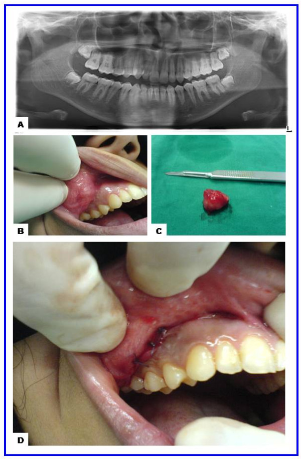
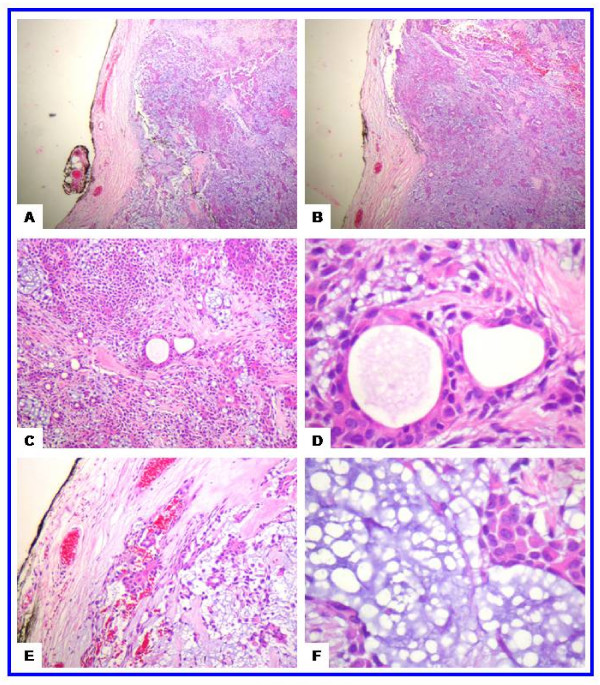
Pleomorphic adenoma, also called benign mixed tumor, is the most common tumor of the salivary glands. About 90% of these tumors occur in the parotid gland and 10% in the minor salivary glands. The most common sites of pleomorphic adenoma of the minor salivary glands are the palates followed by lips and cheeks. Other rare sites include the throat, floor of the mouth, tongue, tonsil, pharynx, retromolar area and nasal cavity. In children, intraoral pleomorphic adenomas of the cheek are extremely rare with only three cases reported to date. Here we report a case of pleomorphic adenoma of minor salivary glands of the cheek in a 17-year-old girl. The mass was removed by wide local excision with adequate margins, and after a follow-up period of three years there were no recurrences. To conclude, pleomorphic adenoma should be considered in the differential diagnosis of cheek masses in youngsters. Wide local excision is to be recommended as the treatment of choice. A close follow-up is necessary postoperatively.


Figures

Figure 1
Figure 1
The radiological and gross features of juvenile pleomorphic adenoma of the cheek. (a) Panoramic radiograph showing unremarkable maxilla, mandible and teeth. (b) Preoperative view showing ovoid nodule in the right cheek covered by intact mucosa with congested blood vessels. (c) Gross features with a well circumscribed ovoid mass measuring 2.0 × 1.7 × 1.5 cm. (d) Postoperative view showing the site of the wound and sutures.
Figure 2
Figure 2
The histological features of the juvenile pleomorphic adenoma of the cheek. (a-b) Scanning magnification views of pleomorphic adenoma showing that the tumor is well circumscribed. The mesenchymal and epithelial elements are evident at these magnifications. This encapsulated tumor has a protuberance pushing through the capsule (upper left side). The tumor has abundant chondroid areas. (c-d) These views show the essential cytoarchitectural features sufficient for diagnosis of pleomoprhic adenoma. The tubular strucutes are well delineated from the stroma. They are lined by inner layer of ductal epithelial cells and other layer of myoepithelial cells. The latter merge imperceptivity into the surrounding mesenchymal elements. The tubules have eosinophilic colloid like materials (secretions) in their lumens. The myoepithelial cells appear as cuboidal, spindle, plasmacytoid and non-descript epithelioid cells. They surround the tubules in a thin layer and in focally thick mantles. (e-f) These views show the stroma (one of the defining component of the pleomorphic adenoma). It is composed of hyaline, homogenous, fibrillary (e) and chondromyoxoid (f) materials interspersed among the epithelial and myoepithelial cells.

References

    1. Garcia Berrocal JR, Ramirez Camacho R, Trinidad A, Salas C. Mixed tumor (pleomorphic adenoma) of head and neck. Typical and atypical patterns. An Otorrinolaringol Ibero Am. 2000;27:333–40. - PubMed
    1. Traiger J, Rosen MB. Mixed Tumor of the Cheek; Report of a Case. Oral Surg Oral Med OralPathol. 1965;19:711–4. doi: 10.1016/0030-4220(65)90339-7. - DOI - PubMed
    1. van Heerden WF, Raubenheimer EJ. Intraoral salivary gland neoplasms: a retrospective study of seventy cases in an African population. Oral Surg Oral Med Oral Pathol. 1991;71:579–82. doi: 10.1016/0030-4220(91)90366-K. - DOI - PubMed
    1. Wang D, Li Y, He H, Liu L, Wu L, He Z. Intraoral minor salivary gland tumors in a Chinese population: a retrospective study on 737 cases. Oral Surg Oral Med Oral Pathol Oral Radiol Endod. 2007;104:94–100. doi: 10.1016/j.tripleo.2006.07.012. - DOI - PubMed
    1. Toida M, Shimokawa K, Makita H, Kato K, Kobayashi A, Kusunoki Y, Hatakeyama D, Fujitsuka H, Yamashita T, Shibata T. Intraoral minor salivary gland tumors: a clinicopathological study of 82 cases. Int J Oral Maxillofac Surg. 2005;34:528–32. doi: 10.1016/j.ijom.2004.10.010. - DOI - PubMed

LinkOut - more resources