. 2009 Oct 15;122(Pt 20):3694-702.
doi: 10.1242/jcs.054700. Epub 2009 Sep 22.

Targeting sequences of UBXD8 and AAM-B reveal that the ER has a direct role in the emergence and regression of lipid droplets

Affiliations

Targeting sequences of UBXD8 and AAM-B reveal that the ER has a direct role in the emergence and regression of lipid droplets

John K Zehmer et al. J Cell Sci. .

Abstract

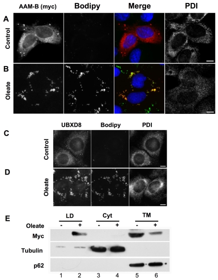
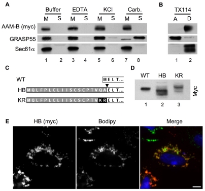
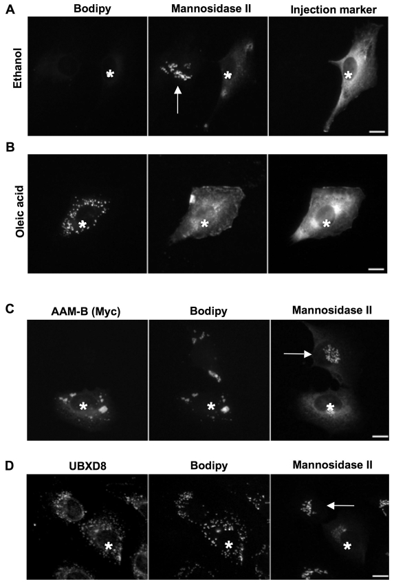
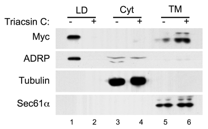
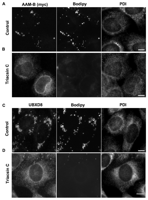
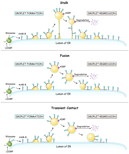
Lipid droplets are sites of neutral lipid storage thought to be actively involved in lipid homeostasis. A popular model proposes that droplets are formed in the endoplasmic reticulum (ER) by a process that begins with the deposition of neutral lipids between the membrane bilayer. As the droplet grows, it becomes surrounded by a monolayer of phospholipid derived from the outer half of the ER membrane, which contains integral membrane proteins anchored by hydrophobic regions. This model predicts that for an integral droplet protein inserted into the outer half of the ER membrane to reach the forming droplet, it must migrate in the plane of the membrane to sites of lipid accumulation. Here, we report the results of experiments that directly test this hypothesis. Using two integral droplet proteins that contain unique hydrophobic targeting sequences (AAM-B and UBXD8), we present evidence that both proteins migrate from their site of insertion in the ER to droplets that are forming in response to fatty acid supplementation. Migration to droplets occurs even when further protein synthesis is inhibited or dominant-negative Sar1 blocks transport to the Golgi complex. Surprisingly, when droplets are induced to disappear from the cell, both proteins return to the ER as the level of neutral lipid declines. These data suggest that integral droplet proteins form from and regress to the ER as part of a cyclic process that does not involve traffic through the secretory pathway.


Figures

Fig. 1.
Fig. 1.
AAM-B and UBXD8 traffic from ER to forming droplets. (A,B) NRK cells on coverslips were transfected with Myc-tagged AAM-B and grown for 8 hours. One set was fixed immediately (A) whereas the other was incubated further in the presence of 50 μg/ml cycloheximide plus oleic acid for 15 hours before fixation (B). The cells were processed for immunofluorescence detection of Myc (red). Neutral lipids were stained with Bodipy 493/503 (green) and DNA with Hoechst 33342 (blue). In uninduced cells, the majority of the staining has a reticular pattern indicative of ER (A). In oleic-acid-treated cells (B) the majority of staining surrounds Bodipy-493/503-positive droplets. Scale bar: 5 μm. (C,D) NRK cells cultured on coverslips were fixed (C) or grown in the presence of cycloheximide plus oleic acid for 15 hours (D). After fixation, cells were processed for immunofluorescence localization of UBXD8 and PDI. Neutral lipids were stained with Bodipy 493/503. In untreated cells, the majority of staining is present in a reticular pattern (C). Oleic acid treatment results in an induction of droplets. As for AAM-B, most of the UBXD8 now surrounds Bodipy-positive droplets (D). Scale bar: 5 μm. (E) CHO K2 cells stably expressing Myc-tagged AAM-B were pretreated with BFA for 8 hours to eliminate droplets. BFA was removed and the cells treated with cycloheximide and either oleic acid or ethanol for 15 hours. The cells were fractionated into droplets (LD), cytosol (Cyt) and total membranes (TM) and equal volumes separated by PAGE and immunoblotted with antibodies against the indicated protein or tag. In ethanol-incubated cells, AAM-B was found exclusively in membranes (lane 5). Incubation in the presence of oleic acid, by contrast, caused a substantial fraction of the AAM-B to move to the droplet fraction (lane 2), with a concomitant loss of AAM-B from the membrane fraction (lane 6). Tubulin and p62 were detected as cytosolic and ER controls, respectively.
Fig. 2.
Fig. 2.
AAM-B is an integral membrane protein made in the ER. Post nuclear supernatants (PNS) prepared from NRK cells transiently expressing Myc-tagged AAM-B were washed with either buffer alone, 10 mM EDTA, 1 M KCl or pH 11 sodium carbonate before centrifugation at 100,000 g. The supernatants and pellets were separated by PAGE and analyzed by immunoblotting. (A) The peripheral membrane protein GRASP55 was extracted from the membranes by carbonate. By contrast, the transmembrane protein Sec61α and AAM-B were found in the membrane pellet under all conditions. (B) Triton-X114 partitioning of the PNS shows that GRASP55 is in the aqueous phase whereas Sec61α and AAM-B partitioned into the detergent phase. (C) Schematic representation of the N-terminal domain of wild-type AAM-B (WT) and two chimeras. The first chimera (HB) has the initiating methionine of AAM-B replaced with the signal sequence from Hepatitis B virus precore protein (highlighted in gray). The predicted signal peptidase cut site is labeled with an arrowhead. The second chimera (KR) is the same as the first but with the Q and A changed to K and R to eliminate the signal sequence cleavage site. (D) HeLa cells were transfected with cDNAs encoding the three constructs, lysates and processed for immunoblotting to detect the Myc tag. The HB construct (lane 2) migrated faster than the WT (lane 1) indicating that the signal sequence had been cleaved. By contrast, the KR construct migrates more slowly than the WT, indicating that the signal sequence has not been cleaved. (E) Cells from the experiment described in D were processed for immunofluorescence detection of Myc-tagged HB using anti-Myc IgG and neutral lipids with Bodipy 493/503. The HB chimera targeted to Bodipy-positive droplets. Scale bar: 5 μm.
Fig. 3.
Fig. 3.
Translocation of AAM-B and UBXD8 from the ER to LDs is independent of Sar1. (A) NRK cells were microinjected with recombinant, dominant-negative Sar1 to block ER exit and cultured for an additional 4 hours before processing for immunofluorescence (asterisk indicates injected cell). Since the Golgi-resident enzyme α-mannosidase II continuously cycles between the ER and Golgi, the block of ER exit in injected cells induces the relocation of α-mannosidase II from the Golgi to the ER. (B) Oleic acid supplementation leads to droplet formation despite the block of ER exit. (C) Cells were co-injected with the dominant-negative Sar1 and cDNA encoding AAM-B. AAM-B is made and trafficked to forming droplets despite the secretory blockade. (D) Endogenous UBXD8 reaches newly formed droplets despite injection of Sar1 dominant-negative protein. Asterisks indicate injected cells. Arrows point to Golgi of uninjected cells. Scale bars: 5 μm.
Fig. 4.
Fig. 4.
AAM-B in droplets returns to a membrane fraction when droplets regress. CHOK2 cells stably expressing Myc-tagged AAM-B were treated with cycloheximide and either triacsin C or DMSO for 15 hours. The cells were fractionated into droplets (LD), cytosol (Cyt) and total membranes (TM) and equal volumes processed for immunoblotting to detect the indicated protein. In control cells, AAM-B-Myc was primarily in droplets (lane 1). Following incubation in the presence of triacsin C, the number of droplets declined as indicated by the loss of ADRP from the LD fraction (lane 2). By contrast, a large increase in the amount of AAM-B in the membrane fraction of the Triacsin C treated cells was observed (lane 6). The ER marker Sec61α remained in the total membrane fraction.
Fig. 5.
Fig. 5.
AAM-B and UBXD8 on LDs return to the ER upon droplet regression. (A,B) NRK cells on coverslips were transfected with Myc-tagged AAM-B. Three hours after transfection, oleate was added and the cells incubated for an additional 6 hours. Cells were then either fixed (A) or incubated in the presence of 1 mg/ml BSA, 50 μg cycloheximide and 7.5 μg/ml triacsin C for 15 hours (B) before processing the samples for immunofluorescence. In the control cells the majority of AAM-B was detected surrounding Bodipy-positive droplets (A). In triacsin-C-treated cells, very few or no droplets were detected by Bodipy staining, and AAM-B was found in a reticular pattern (B). (C,D) NRK cells on coverslips were treated as in A and B, except they were not transfected and the endogenous UBXD8 was detected directly with anti-UBXD8 IgG. In the control cells, UBXD8 was found on Bodipy-positive droplets (C). By contrast, UBXD8 was found primarily in a reticular pattern that colocalized with PDI in the triacsin-C-treated cells (D). Scale bars: 5 μm.
Fig. 6.
Fig. 6.
Three models to explain how integral droplet proteins travel between ER and LD. (A) Stalk Model. LDIMP proteins such as AAM-B and UBXD8 move from the ER to droplets through a stalk composed of two phospholipid monolayers derived from the ER that remains continuous with LDs. During LD regression, LDIMPs migrate back to the ER through the stalk. (B) Fusion Model. LDIMPs move to form droplets that then bud from the ER to form free LDs. During droplet regression, the LD refuses with the ER and the LDIMPs return to the ER. (C) Transient Contact Model. LDIMPs move into droplets that bud from the ER. During droplet regression, the LD docks with the ER outer monolayer where it delivers the L-DIMP back to the ER without fusing.

Similar articles

Cited by

References

    1. Altan-Bonnet, N., Sougrat, R. and Lippincott-Schwartz, J. (2004). Molecular basis for Golgi maintenance and biogenesis. Curr. Opin. Cell Biol. 16, 364-372. - PubMed
    1. Aridor, M., Bannykh, S. I., Rowe, T. and Balch, W. E. (1995). Sequential coupling between copii and copi vesicle coats in endoplasmic reticulum to Golgi transport. J. Cell Biol. 131, 875-893. - PMC - PubMed
    1. Athenstaedt, K., Zweytick, D., Jandrositz, A., Kohlwein, S. D. and Daum, G. (1999). Identification and characterization of major lipid particle proteins of the yeast saccharomyces cerevisiae. J. Bacteriol. 181, 6441-6448. - PMC - PubMed
    1. Bartz, R., Li, W. H., Venables, B., Zehmer, J. K., Roth, M. R., Welti, R., Anderson, R. G., Liu, P. and Chapman, K. D. (2007a). Lipidomics reveals that adiposomes store ether lipids and mediate phospholipid traffic. J. Lipid Res. 48, 837-847. - PubMed
    1. Bartz, R., Seemann, J., Zehmer, J. K., Serrero, G., Chapman, K. D., Anderson, R. G. and Liu, P. (2007b). Evidence that mono-Adp-ribosylation of Ctbp1/bars regulates lipid storage. Mol. Biol. Cell 18, 3015-3025. - PMC - PubMed

Publication types