. 2010 Feb;18(2):413-20.
doi: 10.1038/mt.2009.210. Epub 2009 Sep 22.

Chimeric antigen receptors combining 4-1BB and CD28 signaling domains augment PI3kinase/AKT/Bcl-XL activation and CD8+ T cell-mediated tumor eradication

Affiliations

Chimeric antigen receptors combining 4-1BB and CD28 signaling domains augment PI3kinase/AKT/Bcl-XL activation and CD8+ T cell-mediated tumor eradication

Xiao-Song Zhong et al. Mol Ther. 2010 Feb.

Abstract

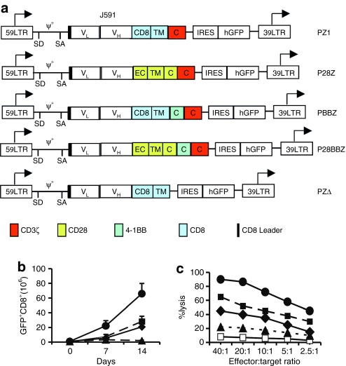
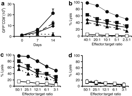
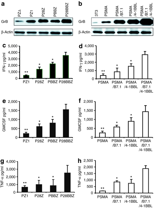
To enhance the strength of activation afforded by tumor antigen-specific receptors, we investigated the effect of adding combined CD28 and 4-1BB costimulatory signaling domains to a chimeric antigen receptor (CAR) specific for prostate-specific membrane antigen (PSMA). Having transferred receptors encompassing the CD28, 4-1BB, and/or CD3zeta cytoplasmic domains in primary human CD8(+) T cells, we find that the P28BBz receptor, which includes all three signaling domains, is superior to receptors that only include one or two of these domains in promoting cytokine release, in vivo T-cell survival and tumor elimination following intravenous T-cell administration to tumor-bearing severe combined immunodeficient (SCID)/beige mice. Upon in vitro exposure to PSMA, the P28BBZ receptor-induced the strongest PI(3)Kinase/Akt activation and Bcl-X(L) expression, and the least apoptosis in transduced peripheral blood CD8(+) T cells. These findings further support the concept of integrating optimized costimulatory properties into recombinant antigen receptors to augment the survival and function of genetically targeted T cells within the tumor microenvironment.


Figures

<b>Figure 1</b>
Figure 1
In vitro proliferation and tumor cytotoxicity of human PSMA-specific primary CD8+ T cells activated with individual or combined CD28 and 4-1BB ligands. (a) Schematic diagram of the Pz1 receptor. The prostate-specific membrane antigen (PSMA)–specific fusion receptors encompass an scFv (white box) derived from the J591 hybridoma, joining the VH and VL fragments through a serine/glycine linker.29 Black: CD8 leader sequence. Red box: CD3ζ chain cytoplasmic domain. Retroviral vector elements: LTR, long terminal repeat; SD, splice donor; SA, splice acceptor; arrows, start of transcription. An EMCV-IRES-humanized renilla green fluorescent protein cassette was cloned downstream of each CAR. (b) Combined CD28 and 4-1BB costimulatory ligands enhance T-cell proliferation and absolute T-cell accumulation following two weekly antigen-specific stimulations. Peripheral blood CD8+ T cells were transduced with Pz1 and activated on AAPCs expressing PSMA and either CD80, CD137, both, or neither. Transduction levels were in the 40–50% range for all receptors (data not shown). T cells were stimulated twice, on day 0 and day 7. Absolute CAR+ T-cell counts are indicated on the y axis. The data represent the mean and SD of nine data points (triplicate cell counts from three separate cultures). (c) Cytolytic activity of antigen-stimulated, CAR-transduced peripheral blood CD8+ T cells. T-cells transduced and activated as described in b were tested on day 7 against RM1.PGLS tumor cells28 and the parental, PSMA RM1. E:T ratios represent the CD8+CAR+ to target cell ratio. Similar results were obtained using EL4PSMA and EL4 cell lines26,29,48 as targets (data not shown). 19z1+ transduced T cells did not lyse PSMA+ tumor cell lines. Closed circles, AAPC-PSMA/B7.1/4-1BBL; closed squares, AAPC-PSMA/B7.1; closed diamond, AAPC-PSMA/4-1BBL; closed triangle, AAPC PSMA; open squares, 19z1 receptor used as negative control.
<b>Figure 2</b>
Figure 2
In vitro proliferation and tumor cytotoxicity of PSMA-targeted human primary CD8+ T cells activated with or without integrated CD28 and 4-1BB costimulation. (a) Integrated CD28 and 4-1BB receptors enhance T-cell proliferation and absolute T-cell accumulation following two weekly antigen-specific stimulations. Peripheral blood CD8+ T cells were transduced with different chimeric antigen receptors (CARs) and subsequently stimulated on the same artificial antigen-presenting cells expressing prostate-specific membrane antigen (PSMA). Transduction levels were in the 40–50% range for all receptors (data not shown). T cells were stimulated twice, on day 0 and day 7. Absolute CAR+ T-cell counts are indicated on the y axis. The data represent the mean and SD of nine data points (triplicate cell counts from three separate cultures). (b) Cytolytic activity of antigen-stimulated, CAR-transduced peripheral blood CD8+ T cells against RM1.PGLS. (c) Cytolytic activity of antigen-stimulated, CAR-transduced peripheral blood CD8+ T cells against the PSMA+ cell line LNCaP. (d) Cytolytic activity of antigen-stimulated, CAR-transduced peripheral blood CD8+ T cells against the PSMA cell line DU145. T cells were transduced, activated, and tested as described in Figure 1. Closed circles, P28BBz; closed squares, P28z; closed diamonds, PBBz; closed triangles, Pz1; open squares, 19z1 receptor (specific for CD19) used as a negative control.
<b>Figure 3</b>
Figure 3
Comparison of the relative potency of prostate-specific membrane antigen-targeted T cells in the RM1.PGLS pulmonary metastases model. Equal numbers of transduced peripheral blood T cells (10 million green fluorescent protein positive CD8+ T cells per mouse) were infused intravenously in tumor-inoculated severe combined immunodeficient/beige mice as previously described.30 Kaplan–Meier survival curves for all treatment groups are represented. Data are pooled from three experiments for a total of 60 mice (Pz1, n = 13; P28z, n = 16; PBBz, n = 13; P28BBz, n = 13 and 19z1, n = 5). Pz1 versus 19z1: P = 0.01; P28BBz versus Pz1: P = 0.025, Log-rank (Mantel–Cox) test.
<b>Figure 4</b>
Figure 4
Integrated 4-1BBL and CD28 costimulation upregulates antigen-elicited Granzyme B, TNF-α, IFN-γ), and granulocyte macrophage–colony stimulating factor (GM-CSF) expression. 1 × 10⁶ chimeric antigen receptor-transduced, rested CD8+ T cells were cocultured with 5 × 10⁵ irradiated cells artificial antigen-presenting cells (AAPCs) expressing either prostate-specific membrane antigen (PSMA), PSMA+B7.1, PSMA+4-1BBL, PSMA+B7.1+4-1BBL or none of the above (“mock,” PSMA AAPC) in the presence or absence of 20 U/ml IL-2. (a,b) Granzyme B expression analyzed by western blot analysis, 24 hours after exposure to antigen. The expression of β-actin was used as a loading control. (a) The Granzyme B expression level in the P28BBz/AAPC-PSMA and (b) Pz1/AAPC-PSMA/B7.1/4-1BBL groups is 3.5- and 6.5-fold greater than the reference Pz1/AAPC-PSMA group, respectively. (c–h) Cytokine expression was analyzed using ELISA or Luminex beads at different time points: IFN-γ (72 hours, c,d), GM-CSF (48 hours, e,f), TNF-α (24 hour, g,h). Error bars indicate standard error (SE) in triplicate samples. *P < 0.05 and **P < 0.001 versus P28BBZ or combination CD28/B7.1 and 4-1BB/4-1BBL signaling.
<b>Figure 5</b>
Figure 5
Integrated 4-1BB and CD28 signals increase in vitro T-cell survival upon antigen restimulation. (a) Inhibition of antigen-induced postactivation cell death. Chimeric antigen receptor (CAR)-transduced CD8+ T cells were restimulated with 3T3-prostate-specific membrane antigen artificial antigen-presenting cells in the presence of 20 U/ml IL-2. After 7 days of coculture, apoptosis was quantified using annexin V and 7-AAD staining. Mean values and SD are from quadruplicate wells from one of three independent experiments performed from different blood donors. *P < 0.05 and **P < 0.001 versus P28BBz. (b) Splenic accumulation of carboxyl fluorescent succinimidyl ester (CFSE)-labeled, CAR-transduced CD8+ T cells 5 days after intravenous adoptive transfer to RM1.PGLS-inoculated severe combined immunodeficient/beige mice (monocistronic vectors encoding CARs only were used in these experiments). (c) Absolute numbers of CD8+ CFSE+ were calculated as the product of percentage transduced cells (determined by flow cytometry) by the total nucleated cell count in the spleen of SCID/beige mice 5 days after adoptive transfer. Each data point represents the average of two or three mice per group. *P < 0.05 and **P < 0.001 versus P28BBz versus CD28/B7.1 and 4-1BB/4-BBL. (d) Cytofluorometric analysis for CFSE-labeled CD8+ T cells 5 days after adoptive transfer, gating on CD3+CD8+ lymphocytes.
<b>Figure 6</b>
Figure 6
Integrated CD28 and 4-1BB–mediated upregulation of Bcl XL is PI3kinase/Akt-dependent. (a) Upregulation of Bcl-XL in P28BBz-transduced, prostate-specific membrane antigen (PSMA)-stimulated CD8+ T cells. Western blot analysis of Bcl-XL expression was performed 24 hours after exposure to PSMA+ artificial antigen-presenting cells (AAPCs). Bcl-XL expression in P28BBz CD8+ T cells is 1.8–3.9-fold greater than with other vectors, as measured by densitometry. (b) Pz1-transduced, rested CD8+ T cells were stimulated with anti-CD3, anti-CD28 and/or anti-4-1BB antibodies in the absence of IL-2. As a control, an isotype-matched IgG was used. Western blot analysis to detect phospho-Akt (ser437) was performed at multiple time points (see Supplementary Figure S2). The 2-hours data shown here are representative of three independent experiments utilizing T cells from three healthy volunteers. (c) P28BBz-transduced CD8+ T cells sustain higher PKB phosphorylation following antigen stimulation. Rested chimeric antigen receptor-transduced CD8+ T cells were cocultured with PSMA-expressing AAPCs without exogenous IL-2, without or with 10 µmol/l Ly29400 or with 0.05% dimethyl sulfoxide for 2 hours. Western blot analysis for phospho-Akt (ser437) and total Akt are shown. Data representative of three independent experiments.

References

    1. Staveley-O'Carroll K, Sotomayor E, Montgomery J, Borrello I, Hwang L, Fein S, et al. Induction of antigen-specific T cell anergy: An early event in the course of tumor progression. Proc Natl Acad Sci USA. 1998;95:1178–1183. - PMC - PubMed
    1. Schwartz RH. T cell anergy. Annu Rev Immunol. 2003;21:305–334. - PubMed
    1. Townsend SE., and , Allison JP. Tumor rejection after direct costimulation of CD8+ T cells by B7-transfected melanoma cells. Science. 1993;259:368–370. - PubMed
    1. Melero I, Bach N, Hellström KE, Aruffo A, Mittler RS., and , Chen L. Amplification of tumor immunity by gene transfer of the co-stimulatory 4-1BB ligand: synergy with the CD28 co-stimulatory pathway. Eur J Immunol. 1998;28:1116–1121. - PubMed
    1. Weinberg AD, Rivera MM, Prell R, Morris A, Ramstad T, Vetto JT, et al. Engagement of the OX-40 receptor in vivo enhances antitumor immunity. J Immunol. 2000;164:2160–2169. - PubMed

Publication types

MeSH terms

Substances