

Differential transduction following basal ganglia administration of distinct pseudotyped AAV capsid serotypes in nonhuman primates
- PMID: 19773746
- PMCID: PMC2839447
- DOI: 10.1038/mt.2009.216
Differential transduction following basal ganglia administration of distinct pseudotyped AAV capsid serotypes in nonhuman primates
Abstract
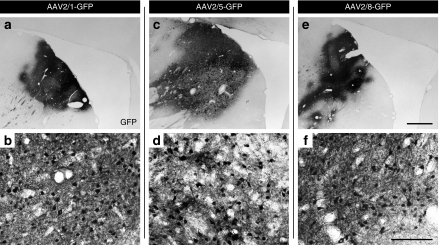
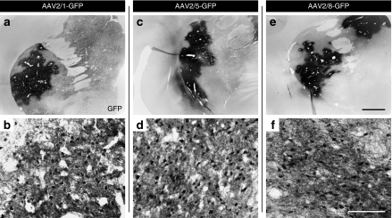
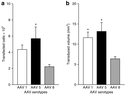
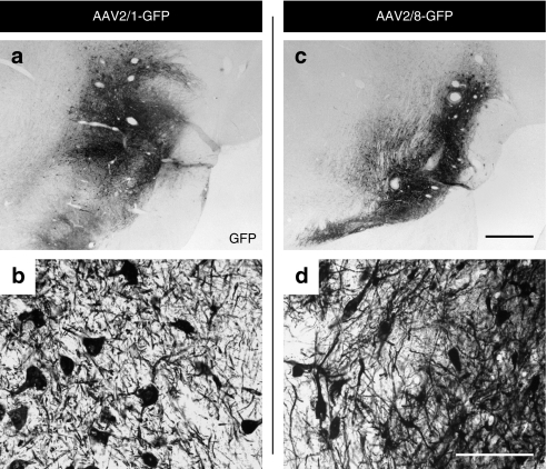
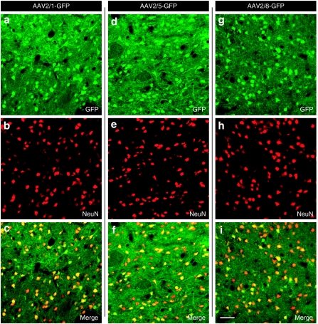
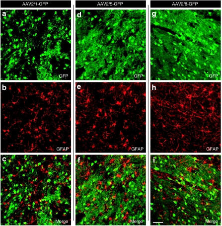
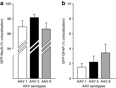
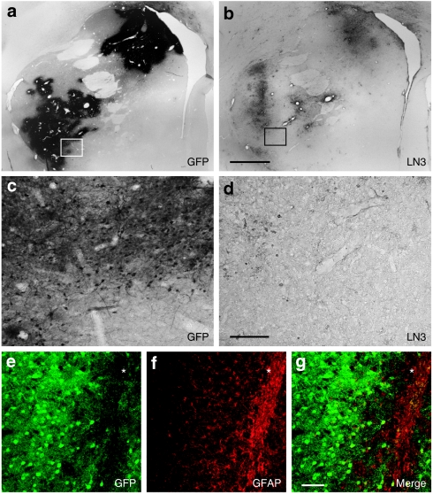
We examined the transduction efficiency of different adeno-associated virus (AAV) capsid serotypes encoding for green fluorescent protein (GFP) flanked by AAV2 inverted terminal repeats in the nonhuman primate basal ganglia as a prelude to translational studies, as well as clinical trials in patients with Parkinson's disease (PD). Six intact young adult cynomolgus monkeys received a single 10 microl injection of AAV2/1-GFP, AAV2/5-GFP, or AAV2/8-GFP pseudotyped vectors into the caudate nucleus and putamen bilaterally in a pattern that resulted in each capsid serotype being injected into at least four striatal sites. GFP immunohistochemistry revealed excellent transduction rates for each AAV pseudotype. Stereological estimates of GFP+ cells within the striatum revealed that AAV2/5-GFP transduces significantly higher number of cells than AAV2/8-GFP (P < 0.05) and there was no significant difference between AAV2/5-GFP and AAV2/1-GFP (P = 0.348). Consistent with this result, Cavalieri estimates revealed that AAV2/5-GFP resulted in a significantly larger transduction volume than AAV2/8-GFP (P < 0.05). Each pseudotype transduced striatal neurons effectively [>95% GFP+ cells colocalized neuron-specific nuclear protein (NeuN)]. The current data suggest that AAV2/5 and AAV2/1 are superior to AAV2/8 for gene delivery to the nonhuman primate striatum and therefore better candidates for therapeutic applications targeting this structure.
Figures
Similar articles
-
Adeno-associated viral serotypes differentially transduce inhibitory neurons within the rat amygdala.Brain Res. 2017 Oct 1;1672:148-162. doi: 10.1016/j.brainres.2017.07.023. Epub 2017 Jul 29. Brain Res. 2017. PMID: 28764932
-
Comparative transduction efficiency of AAV vector serotypes 1-6 in the substantia nigra and striatum of the primate brain.Mol Ther. 2010 Mar;18(3):588-93. doi: 10.1038/mt.2009.286. Epub 2009 Dec 15. Mol Ther. 2010. PMID: 20010918 Free PMC article.
-
Recombinant adeno-associated virus type 2 pseudotypes: comparing safety, specificity, and transduction efficiency in the primate striatum. Laboratory investigation.J Neurosurg. 2011 Mar;114(3):672-80. doi: 10.3171/2010.8.JNS091583. Epub 2010 Oct 15. J Neurosurg. 2011. PMID: 20950087 Free PMC article.
-
Glial-derived neurotrophic factor gene transfer for Parkinson's disease: anterograde distribution of AAV2 vectors in the primate brain.Neurobiol Dis. 2012 Nov;48(2):228-35. doi: 10.1016/j.nbd.2011.10.004. Epub 2011 Oct 14. Neurobiol Dis. 2012. PMID: 22019719 Free PMC article. Review.
-
Advances in AAV technology for delivering genetically encoded cargo to the nonhuman primate nervous system.Curr Res Neurobiol. 2023 Apr 7;4:100086. doi: 10.1016/j.crneur.2023.100086. eCollection 2023. Curr Res Neurobiol. 2023. PMID: 37397806 Free PMC article. Review.
Cited by
-
Impact of age and vector construct on striatal and nigral transgene expression.Mol Ther Methods Clin Dev. 2016 Dec 7;3:16082. doi: 10.1038/mtm.2016.82. eCollection 2016. Mol Ther Methods Clin Dev. 2016. PMID: 27933309 Free PMC article.
-
Gene therapy for Parkinson's disease.Parkinsons Dis. 2012;2012:757305. doi: 10.1155/2012/757305. Epub 2012 Mar 25. Parkinsons Dis. 2012. PMID: 22619738 Free PMC article.
-
Optogenetics in the nonhuman primate.Prog Brain Res. 2012;196:215-33. doi: 10.1016/B978-0-444-59426-6.00011-2. Prog Brain Res. 2012. PMID: 22341328 Free PMC article. Review.
-
Adeno-Associated Virus Technologies and Methods for Targeted Neuronal Manipulation.Front Neuroanat. 2019 Nov 26;13:93. doi: 10.3389/fnana.2019.00093. eCollection 2019. Front Neuroanat. 2019. PMID: 31849618 Free PMC article. Review.
-
Early or late-stage anti-N-terminal Huntingtin intrabody gene therapy reduces pathological features in B6.HDR6/1 mice.J Neuropathol Exp Neurol. 2010 Oct;69(10):1078-85. doi: 10.1097/NEN.0b013e3181f530ec. J Neuropathol Exp Neurol. 2010. PMID: 20838238 Free PMC article.
References
-
- Muzyczka N. Use of adeno-associated virus as a general transduction vector for mammalian cells. Curr Top Microbiol Immunol. 1992;158:97–129. - PubMed
-
- Hildinger M., and , Auricchio A. Advances in AAV-mediated gene transfer for the treatment of inherited disorders. Eur J Hum Genet. 2004;12:263–271. - PubMed
-
- Bartlett JS, Samulski RJ., and , McCown TJ. Selective and rapid uptake of adeno-associated virus type 2 in brain. Hum Gene Ther. 1998;9:1181–1186. - PubMed
-
- Xu R, Janson CG, Mastakov M, Lawlor P, Young D, Mouravlev A, et al. Quantitative comparison of expression with adeno-associated virus (AAV-2) brain-specific gene cassettes. Gene Ther. 2001;8:1323–1332. - PubMed
Publication types
MeSH terms
Substances
Grants and funding
LinkOut - more resources
Full Text Sources
Other Literature Sources
Medical
