

Direct cancer-stromal interaction increases fibroblast proliferation and enhances invasive properties of scirrhous-type gastric carcinoma cells
- PMID: 19773759
- PMCID: PMC2768433
- DOI: 10.1038/sj.bjc.6605309
Direct cancer-stromal interaction increases fibroblast proliferation and enhances invasive properties of scirrhous-type gastric carcinoma cells
Abstract
Background: Scirrhous-type gastric carcinoma (SGC) exhibits an extensive submucosal fibrosis and extremely poor patient prognosis. We investigated the importance of the cancer-stromal interaction in the histogenesis of SGC.
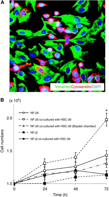
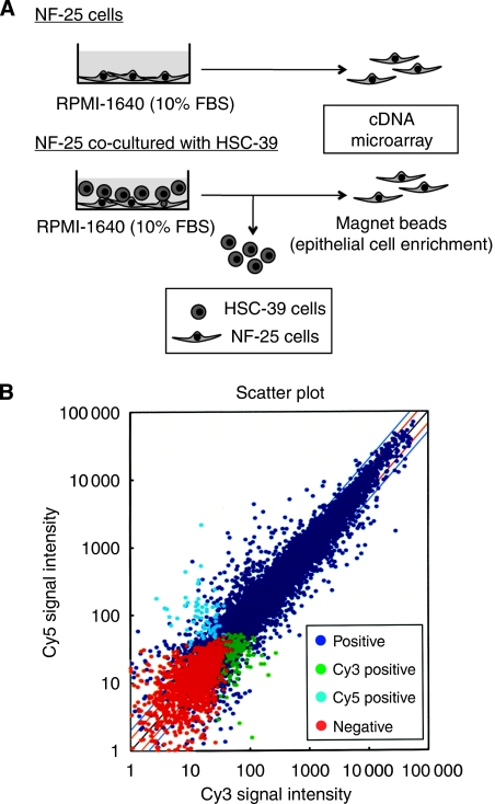
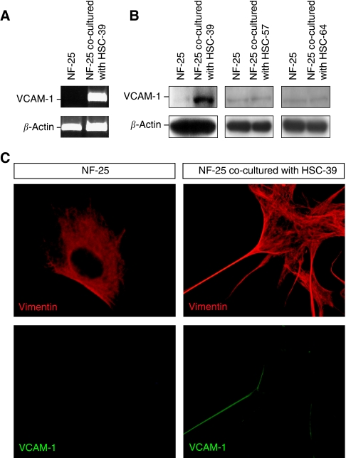
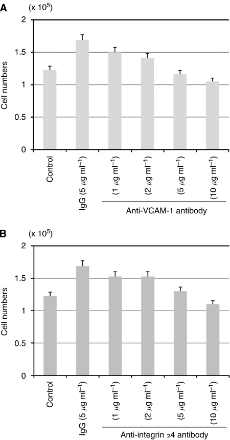
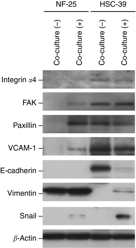
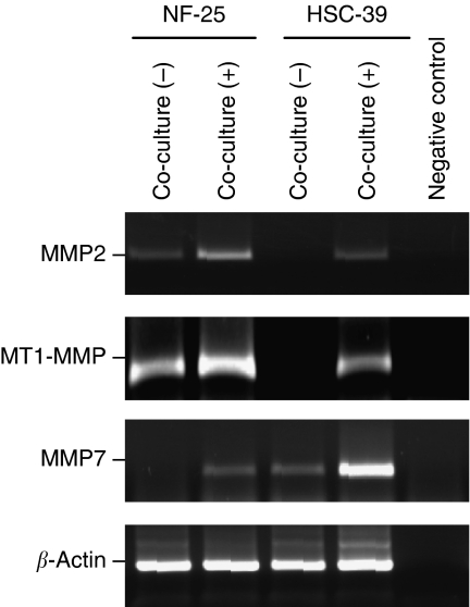
Methods: Gastric fibroblasts NF-25 and intestinal fibroblasts NF-j2 were co-cultured with SGC-derived (HSC-39) or non-SGC-derived (HSC-57 and HSC-64) cells. To identify genes that are up- or downregulated in NF-25, complementary DNA (cDNA) microarray analysis was performed. The antibody against vascular-cell adhesion molecule-1 (VCAM-1) was used for cell growth test and immunohistochemistry. Moreover, the impact of interaction with NF-25 fibroblasts on HSC-39 cells was investigated using western blot and reverse transcription-polymerase chain reaction.
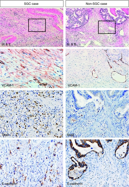
Results: HSC-39 cells stimulated growth of NF-25 but not NF-j2 when co-cultured. Induction of VCAM-1 in NF-25 fibroblasts was identified, which was specific when co-cultured with HSC-39 but not with non-SGC-derived HSC-57 and HSC-64 cells. Neutralising antibody to VCAM-1 suppressed NF-25 growth in dose-dependent manners. In tissue samples, positive immunoreactivity of VCAM-1 in SGC-derived fibroblasts was significantly higher than that in non-SGC-derived fibroblasts. Furthermore, interaction with NF-25 fibroblasts not only induced the epithelial-mesenchymal transition-like change, but also expressions of matrix metalloproteinase- related genes in HSC-39 cells.
Conclusion: Direct interaction between SGC cells and gastric fibroblasts establishes the tumour microenvironment and reinforces the aggressiveness of SGC.
Figures
Similar articles
-
Mesothelial cells differentiate into fibroblast-like cells under the scirrhous gastric cancer microenvironment and promote peritoneal carcinomatosis in vitro and in vivo.Mol Cell Biochem. 2013 May;377(1-2):177-85. doi: 10.1007/s11010-013-1583-0. Epub 2013 Feb 8. Mol Cell Biochem. 2013. Retraction in: Mol Cell Biochem. 2024 Jan;479(1):197-198. doi: 10.1007/s11010-023-04911-z. PMID: 23392771 Retracted.
-
Stromal fibroblasts mediate extracellular matrix remodeling and invasion of scirrhous gastric carcinoma cells.PLoS One. 2014 Jan 10;9(1):e85485. doi: 10.1371/journal.pone.0085485. eCollection 2014. PLoS One. 2014. PMID: 24427313 Free PMC article.
-
Selective cyclooxygenase-2 inhibitor downregulates the paracrine epithelial-mesenchymal interactions of growth in scirrhous gastric carcinoma.Int J Cancer. 2007 Feb 1;120(3):686-93. doi: 10.1002/ijc.22329. Int J Cancer. 2007. PMID: 17096355
-
The significance of scirrhous gastric cancer cell lines: the molecular characterization using cell lines and mouse models.Hum Cell. 2018 Oct;31(4):271-281. doi: 10.1007/s13577-018-0211-4. Epub 2018 Jun 6. Hum Cell. 2018. PMID: 29876827 Review.
-
[Pathogenesis and progression of scirrhous carcinoma].Gan To Kagaku Ryoho. 1994 Oct;21(14):2364-70. Gan To Kagaku Ryoho. 1994. PMID: 7944478 Review. Japanese.
Cited by
-
Cancer-associated fibroblasts regulate the biological behavior of cancer cells and stroma in gastric cancer.Oncol Lett. 2018 Jan;15(1):691-698. doi: 10.3892/ol.2017.7385. Epub 2017 Nov 9. Oncol Lett. 2018. PMID: 29399141 Free PMC article.
-
Intratumor stromal proportion predicts aggressive phenotype of gastric signet ring cell carcinomas.Gastric Cancer. 2017 Jul;20(4):591-601. doi: 10.1007/s10120-016-0669-2. Epub 2016 Nov 17. Gastric Cancer. 2017. PMID: 27858181
-
Potential biomarkers and signaling pathways associated with the pathogenesis of primary salivary gland carcinoma: a bioinformatics study.Genomics Inform. 2021 Dec;19(4):e42. doi: 10.5808/gi.21052. Epub 2021 Dec 31. Genomics Inform. 2021. PMID: 35012286 Free PMC article.
-
Stromal fibroblasts in the microenvironment of gastric carcinomas promote tumor metastasis via upregulating TAGLN expression.BMC Cell Biol. 2013 Mar 19;14:17. doi: 10.1186/1471-2121-14-17. BMC Cell Biol. 2013. PMID: 23510049 Free PMC article.
-
Notch and TGF-β/Smad3 pathways are involved in the interaction between cancer cells and cancer-associated fibroblasts in papillary thyroid carcinoma.Tumour Biol. 2014 Jan;35(1):379-85. doi: 10.1007/s13277-013-1053-z. Epub 2013 Aug 6. Tumour Biol. 2014. PMID: 23918305
References
-
- Alexiou D, Karayiannakis AJ, Syrigos KN, Zbar A, Kremmyda A, Bramis I, Tsigris C (2002) Serum levels of E-selectin, ICAM-1 and VCAM-1 in colorectal cancer patients: corrrelations with clinicopathological features, patient survival and tumour surgery. Eur J Cancer 37: 2392–2397 - PubMed
-
- Berx G, Raspe E, Christofori G, Thiery JP, Sleeman JP (2007) Pre-EMTing metastasis? Recapitulation of morphogenetic processes in cancer. Clin Exp Metastasis 24: 587–597 - PubMed
-
- Bogetto L, Gabriele E, Cariati R, Dolcetti R, Spessotto P, Doglioni C, Boiocchi M, Perris R, Colombatti A (2000) Bidirectional induction of the cognate receptor–ligand α4/VCAM-1 pair defines a novel mechanism of tumor intravasation. Blood 95: 2397–2406 - PubMed
-
- Chan PY, Aruffo A (1993) VLA-r integrin mediates lymphocyte migration on the inducible endothelial ligand VCAM-1 and the extracellular matrix protein fibronectin. J Biol Chem 268: 246555–246564 - PubMed
-
- Cui W, Fowlis DJ, Bryson S, Duffie E, Ireland H, Balmain A, Akhurst RJ (1996) TGFβ1 inhibits the formation of benign skin tumors, but enhances progression to invasive spindle carcinomas in transgenic mice. Cell 86: 531–542 - PubMed
Publication types
MeSH terms
Substances
LinkOut - more resources
Full Text Sources
Medical
Miscellaneous
