. 2009 Feb;19(1):69-74.
doi: 10.4103/0971-3026.45349.

Pictorial essay: MRI of the fetal brain

Pictorial essay: MRI of the fetal brain

Ganesh Rao B et al. Indian J Radiol Imaging. 2009 Feb.

Abstract

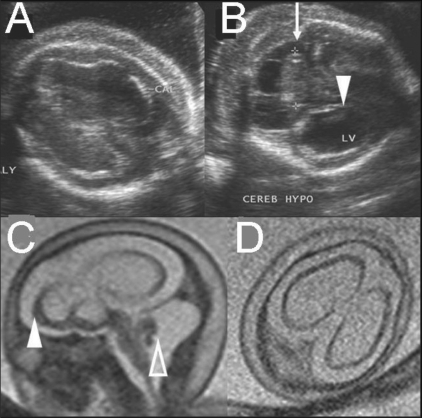
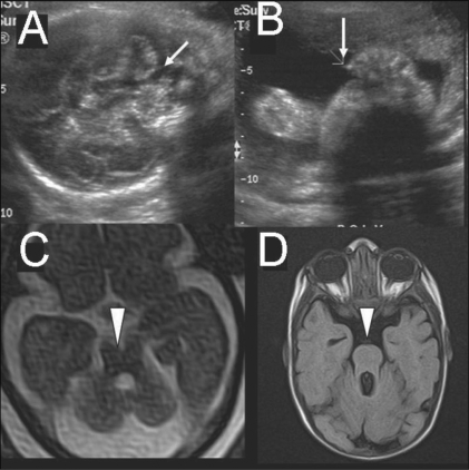
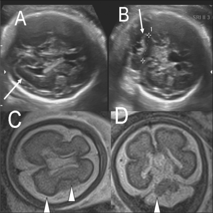
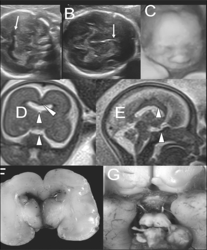
MRI is a useful supplement to USG for the assessment of fetal brain malformations. Superior soft tissue contrast and the ability to depict sulcation and myelination are the strengths of MRI. Subtle or inconclusive USG abnormalities can be confirmed or ruled out by MRI. In some cases, additional findings detected with MRI often help in arriving at a definitive diagnosis, which is necessary for parental counseling and for guiding management. Fast T2W sequences form the basis of fetal MRI. There have been no reports of deleterious effects of MRI on the fetus. A few case examples are presented to illustrate the advantages of MRI.

Keywords: Brain; MRI; fetal.


Conflict of interest statement

Conflict of Interest: None declared.

Figures

Figure 1 (A,B)
Figure 1 (A,B)
T2W sagittal MRI image (B) at 27 weeks shows the parieto-occipital sulcus (arrowhead), calcarine sulcus (open arrow), and primary cerebellar fissure (closed arrow). A T2W axial MRI image (B) shows myelination (hypointense region) in the posterior brainstem (arrowhead)
Figure 2 (A,B)
Figure 2 (A,B)
Single live gestation at 33 weeks. T2W (A) and gradient-echo T2W (T2*) MRI images show intraventricular (arrows) and periventricular (arrowhead) hemorrhage. Fetal blood sampling was negative for TORCH IgMs. A platelet count of 50,000 / mm3 was noted. Pregnancy ended with a stillbirth at 35 weeks.
Figure 3
Figure 3
Single live gestation at 30 weeks. T1W axial MRI image at the level of the lateral ventricles shows tubers as subependymal hyperintense nodules (arrows).
Figure 4 (A-D)
Figure 4 (A-D)
Single live gestation at 37 weeks. USG scans of the heart (A) and brain (B) show a cardiac rhabdomyoma (arrow in A) and mild ventriculomegaly (arrow in B). Axial (C) and coronal (D) T2W MRI images show subependymal tubers (arrowheads), confirming the diagnosis of tuberous sclerosis. The large left lateral ventricular subependymal nodule at the foramen of Monroe is probably responsible for the unilateral ventriculomegaly. The infant developed ash-leaf macules at 1 month and myoclonic jerks at 2 months of age. It died at 3 months (cradle death)
Figure 5 (A-F)
Figure 5 (A-F)
Single live gestation at 24 weeks. USG images (A-C) in different planes show ventriculomegaly with prominent occipital horns (arrows) and widened subarachnoid spaces. The possibilities of neuronal migrational and an atrophic disorder were considered. T2W axial (D), coronal (E), and sagittal (F) MRI images show bilateral symmetric total neuroparenchymal loss in the parieto-occipital watershed regions (arrowheads) but no clefting; this is suggestive of an ischemic etiopathogenesis rather than a neuronal migrational disorder. The couple opted for termination of pregnancy. They declined an autopsy
Figure 6 (A-D)
Figure 6 (A-D)
Single live gestation at 25 weeks. USG images (A,B) show cortical atrophy (A) and cerebellar hypoplasia (arrow in B), ventriculomegaly, and periventricular calcification (arrowhead in B). Sagittal T2W MRI image (C) shows marked cortical thinning (solid arrowhead) and marked cerebellar hypoplasia (open arrowhead) in a suspected case of fetal CMV infection. Axial T2W MRI image (D) fails to demonstrate calcification. The couple declined prenatal testing and opted for termination of pregnancy.
Figure 7 (A-D)
Figure 7 (A-D)
Single live gestation at 28 weeks. USG images (A,B) show inferior vermian agenesis (arrow in A) and postaxial polydactyly (arrow in B). T1W axial image (C) does not show the ‘molar tooth sign’ (arrowhead). Postnatal T1W axial MRI image at 4 months shows the ‘molar tooth sign’ (arrowhead), confirming the diagnosis of Joubert's syndrome. In this case, the mother noticed abnormal eye movements and failure of fixation of vision by the infant at 3 months of age
Figure 8 (A-D)
Figure 8 (A-D)
Single live gestation at 31 weeks. USG images (A,B) show suspected cortical atrophy (arrow in A) and cerebellar hypoplasia (arrow in B). Axial T2W HASTE MRI image (C) shows poor sulcation and shallow Sylvian fissures with poor opercularization (arrowheads), diagnostic of lissencephaly. Coronal T2W MRI image (D) shows cerebellar hypoplasia (arrowhead). This case was lost to follow-up
Figure 9 (A-G)
Figure 9 (A-G)
Pregestational diabetic mother with a 20 weeks' single live gestation. USG images (A,B) show fusion of the frontal horns of the lateral ventricles (arrow in A) and absent cavum septum pellucidum (arrow in B), indicating callosal agenesis. 3-D USG image (C) shows a normal fetal face. T2W coronal MRI image (D) shows absent septum pellucidum, midline cerebral fusion (solid arrowhead), and indistinct optic chiasm (open arrowhead). T2W sagittal MRI image shows the presence of a corpus callosum (open arrowhead) but a possibly hypoplastic hypophysis (solid arrowhead). Autopsy (F,G) images show frontal horn fusion, absent septum pellucidum, and a small optic chiasm, confirming the diagnosis of septo-optic dysplasia

Similar articles

References

    1. Levine D, Barnes D, Madsen JR, Li W, Edelman RR. Fetal central nervous system anomalies: MR imaging augments sonographic diagnosis. Radiology. 1997;204:35–64. - PubMed
    1. Coakley FV, Glenn OA, Qayyum A, Barkovich AJ, Golstein R, Filly RA. Fetal MRI: A developing technique for the developing patient. AJR Am J Roentgenol. 2004;182:242–53,55. - PubMed
    1. Twickler DM, Magee KP, Caire J, Zaretsky M, Fleckenstein JL, Ramus RM. Second-opinion magnestic resonance imaging for suspected fetal central nervous system abnormalities. Am J Obstet Gynecol. 2003;188:492–6. - PubMed
    1. Sonigo C, Rypens FF, Carteret M, Delezoide AL, Brunelle FO. MR imaging of fetal cerebral anomalies. Pediatr Radiol. 1998;28:212–22. - PubMed
    1. Garel C. Berlin: Springer; 2004. MRI of the Fetal Brain; p. 267.