Case Report: Congenital infiltrating lipomatosis of face
- PMID: 19774187
- PMCID: PMC2747453
- DOI: 10.4103/0971-3026.43847
Case Report: Congenital infiltrating lipomatosis of face
Abstract
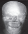
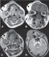
Congenital infiltrating lipomatosis of the face is a rare condition characterized by diffuse fatty infiltration of the facial soft tissues. There may be muscle involvement along with associated bony hyperplasia. It is a type of lipomatous tumor that is congenital in origin; it is rare and seen usually in childhood. We recently saw an 11-year-old girl with this condition. She presented with a swelling of the right side of the face that had been present since birth; there were typical findings on plain radiographs, CT, and MRI. The patient underwent cosmetic surgery. Histopathological examination showed mature adipocytes without any capsule.
Keywords: Congenital; lipomatosis.
Conflict of interest statement
Figures


References
-
- Slavin SA, Baker DC, McCarthy JG, Muffarij A. Congenital infiltrating lipomatosis of the face: Clinicopathologic evaluation and treatment. Plast Reconstr Surg. 1983;72:158–64. - PubMed
-
- Heymans O, Ronsmans C. Congenital infiltrating lipomatosis of the face. Eur J Plast Surg. 2005;28:186–9.
-
- Haloi AK, Ditchfield M, Penington A, Phillips R. Facial infiltrative lipomatosis. Pediatr Radiol. 2006;36:1159–62. - PubMed
-
- Chen CM, Lo LJ, Wong HF. Congenital infiltrating lipomatosis of the face: Case report and literature review. Chang Gung Med J. 2002;25:194–200. - PubMed
-
- Coffin CM. A clinical, pathological, and therapeutic approach. Baltimore: Williams and Wilkins Co; 1997. Adipose and myxoid tumors. Pediatric soft tissue tumors; p. 254.
LinkOut - more resources
Full Text Sources

