. 2010 Jan;222(1):103-10.
doi: 10.1002/jcp.21925.

Human embryonic stem cells are pre-mitotically committed to self-renewal and acquire a lengthened G1 phase upon lineage programming

Affiliations

Human embryonic stem cells are pre-mitotically committed to self-renewal and acquire a lengthened G1 phase upon lineage programming

Klaus A Becker et al. J Cell Physiol. 2010 Jan.

Abstract

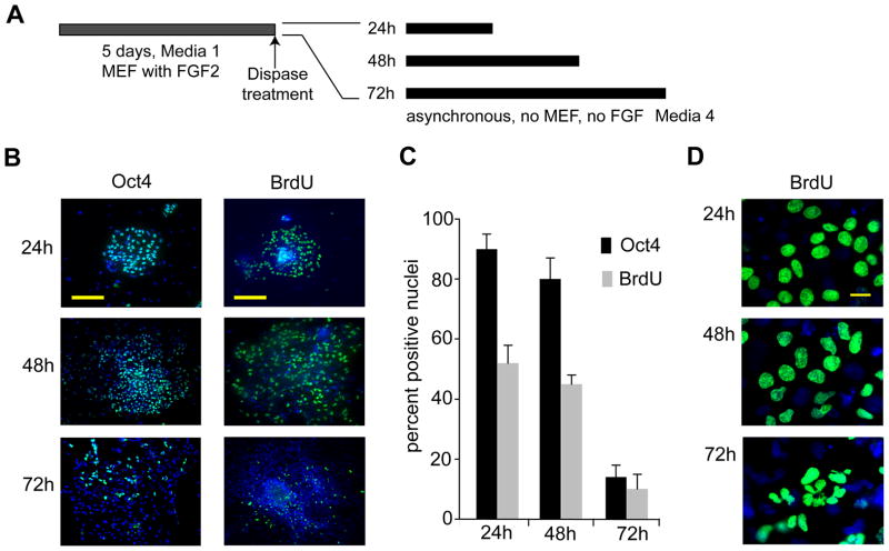
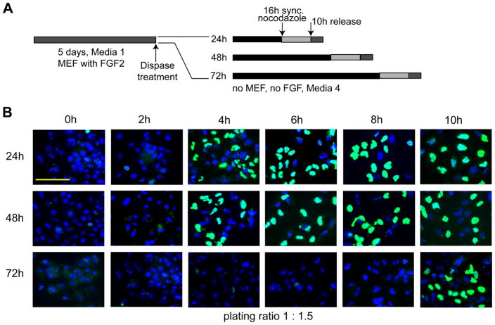
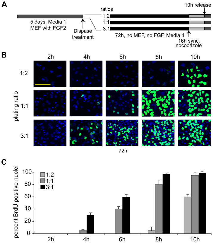
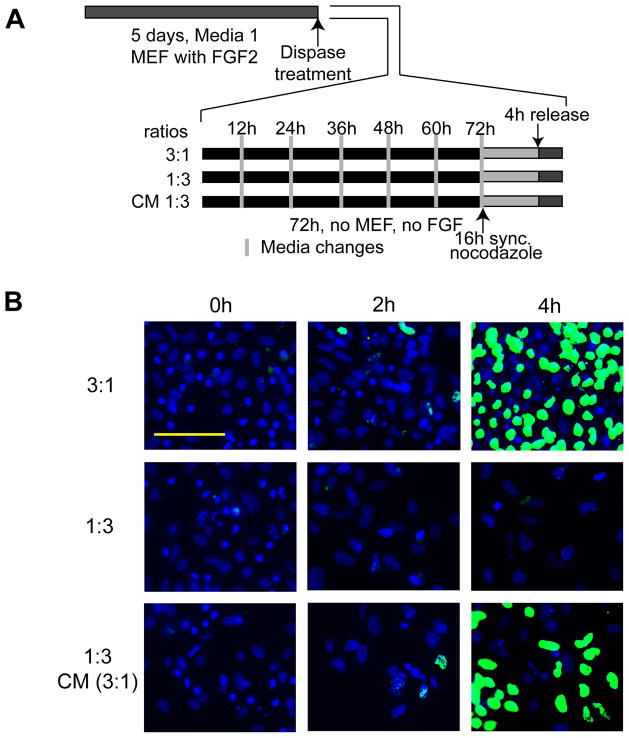
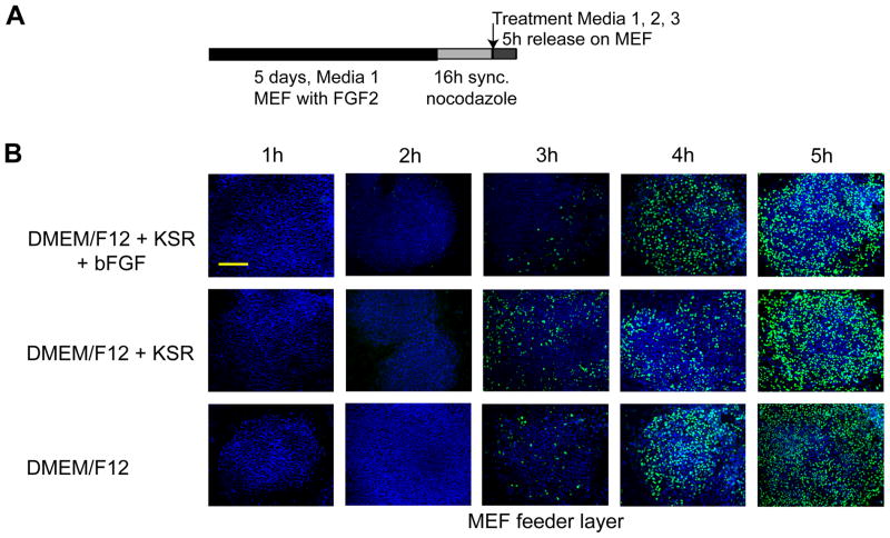
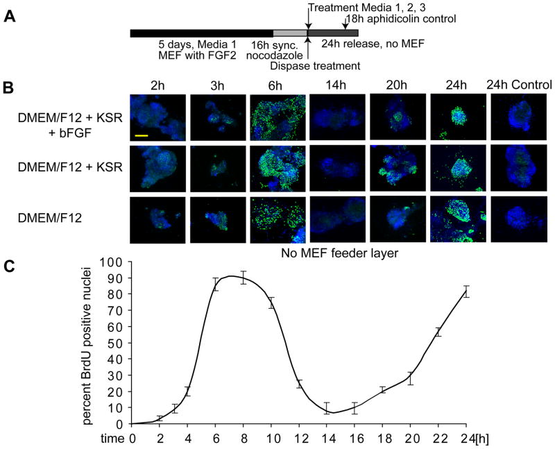
Self-renewal of human embryonic stem (hES) cells proceeds by a unique abbreviated cell cycle with a shortened G1 phase and distinctions in molecular cell cycle regulatory parameters. In this study, we show that early lineage-commitment of pluripotent hES cells modifies cell cycle kinetics. Human ES cells acquire a lengthened G1 within 72 h after lineage-programming is initiated, as reflected by loss of the pluripotency factor Oct4 and alterations in nuclear morphology. In hES cells that maintain the pristine pluripotent state, we find that autocrine mechanisms contribute to sustaining the abbreviated cell cycle. Our data show that naïve and mitotically synchronized pluripotent hES cells are competent to initiate two consecutive S phases in the absence of external growth factors. We conclude that short-term self-renewal of pluripotent hES cells occurs autonomously, in part due to secreted factors, and that pluripotency is functionally linked to the abbreviated hES cell cycle.


Figures

Figure 1
Figure 1. Reduced DNA synthesis in hES cells is linked to loss of pluripotency
(A) Experimental design. (B) Immunofluorescence microscopy detection of Oct4 and BrdU in asynchronous human ES cells (WA09/H9) at 24 h, 48 h, and 72 h and cultured in differentiating medium under feeder-free conditions (scale bar = 200 μm). (C) Quantitation of results depicted in (B); the error-bars represent the standard error of the mean (SEM) for four independent determinations of 400 each (n=1600). Changes in Oct4 and BrdU signals between 48h and 72 h are statistically significant (P<0.05) (D) Micrographs of nuclei at higher magnification show changes in nuclear morphology after 24 h 48 h, and 72 h under differentiating conditions (scale bar = 20 μm).
Figure 2
Figure 2. Extension of the G1 phase in human ES cells during early lineage-programming
(A) Experimental design. (B) Human ES cells were cultured in differentiating media for 24 h, 48 h, and 72 h under feeder-free conditions (first, second and third rows, respectively). Cell cultures were then synchronized using nocodazole for an additional 16 h and subsequently released. BrdU incorporation in nuclei was detected by immunofluorescent microscopy (scale bar = 100 μm) using cells taken at the time of release (0 h) and at 2 h intervals for up to 10 h (first through sixth column, respectively). Cells were plated at a 1:1.5 split ratio (see Figure 3).
Figure 3
Figure 3. Lengthening of the G1 phase in human ES cells depends on cell density
(A) Experimental design. (B) Human ES cells were seeded in differentiating media under feeder-free and conditions at low (1:2), medium (1:1), and high density (3:1) for 72 h. These ratios reflect the split of the original culture. In a split of 1:2, colonies from one plate were divided into two, a 3:1 ratio means that colonies from three plates were pooled into one plate. At 72 h, cells were mitotically arrested using nocodazole for an additional 16 h, and cell cycle progression was monitored using immunofluorescence detection of BrdU incorporation at 2 h, 4 h, 6 h, 8 h, and 10 h after mitotic release (Scale bar = 100 μm). (C) Quantitation of the fraction of BrdU labeled nuclei at the indicated time points after culture at different plating densities (as reflected by split ratios). The quantitation is based on two independent experiments performed in duplicate in which 400 nuclei were counted (n=1600; error bars = SEM).
Figure 4
Figure 4. Medium conditioned by hES cells prevents lengthening of the G1 phase
(A) Experimental design. (B) Human ES cells were plated without feeder layer at high density (3:1) (first row) and low density (1:3) (second and third rows), and maintained with differentiation media for 72 h. Media changes were applied every 12 h. Control cells grown in high density (first row) or low density (second row) received fresh media, but cells in grown in low density (1:3) (third row) received the conditioned medium (CM) supernatant of cells grown in high density (3:1, first row) every 12 h. After 72 h, cultures were synchronized for an additional 16 h using nocodazole. Upon release from mitotic block, BrdU incorporation was determined at 0 h, 2 h and 4 h to determine the duration of G1 and the onset of S phase (scale bar = 100 μm).
Figure 5
Figure 5. G1 phase progression in human ES cells is independent of exogenous FGF2
(A) Experimental design. (B) Mitotically synchronized WA09/H9 cells were generated in standard hES medium (Medium 1) on a feeder layer of inactivated mouse embryo fibroblasts (MEFs) by mitotic inhibition using nocodazole for 16 h. Cells were then released from mitotic block and allowed to reenter the cell cycle in the presence of fresh complete DMEM/F12 medium (Medium 1) (first row), the same medium but without bFGF/FGF2 (Medium 2) (second row), or incomplete DMEM/F12 medium (Media 3) (third row). Cell cycle progression was examined at the indicated hourly time points after release (columns) by monitoring BrdU incorporation (scale bar = 200 μm).
Figure 6
Figure 6. Human ES cells have the intrinsic ability to enter two consecutive S phases
(A) Experimental design. (B) Human ES cells were synchronized using nocodazole and mitotically blocked colonies were separated from the inactivated MEF feeder layer by gentle mechanical disruption and dispase treatment. Colonies were then seeded on plastic coverslips (i.e., without feeder layer) and cultures were supplemented under different culture conditions: regular human ES cell media (Medium 1) (first row), medium in which FGF2 is omitted (Medium 2) (second row) and medium consisting of DMEM/F12 only (Medium 3) (third row). After adherence (< 1 h), BrdU incorporation was determined for multiple time-points at increasing temporal intervals as indicated (columns) to cover cell cycle progression through two consecutive cell cycles (up to 24 h). In parallel, we treated a control group of cells at18 h after mitotic release (i.e., after one cell cycle) with aphidicolin to prevent the initiation of a second S phase (24 h Control) (scale bar = 200 μm). The bottom panel shows a line graph that quantifies BrdU incorporation as a function of time after mitotic release and reflects the oscillating behavior expected from progression into two consecutive S phases. (C) Shows the quantification of results depicted in (B); the error-bars represent SEM for four independent determinations of 400 each (n=1600).

Similar articles

Cited by

References

    1. Amit M, Carpenter MK, Inokuma MS, Chiu CP, Harris CP, Waknitz MA, Itskovitz-Eldor J, Thomson JA. Clonally derived human embryonic stem cell lines maintain pluripotency and proliferative potential for prolonged periods of culture. Dev Biol. 2000;227:271–278. - PubMed
    1. Becker KA, Ghule PN, Therrien JA, Lian JB, Stein JL, van Wijnen AJ, Stein GS. Self-renewal of human embryonic stem cells is supported by a shortened G1 cell cycle phase. J Cell Physiol. 2006;209:883–893. - PubMed
    1. Becker KA, Stein JL, Lian JB, van Wijnen AJ, Stein GS. Establishment of histone gene regulation and cell cycle checkpoint control in human embryonic stem cells. J Cell Physiol. 2007;210:517–526. - PubMed
    1. Blagosklonny MV, Pardee AB. The restriction point of the cell cycle. Cell Cycle. 2002;1:103–110. - PubMed
    1. Bodnar MS, Meneses JJ, Rodriguez RT, Firpo MT. Propagation and maintenance of undifferentiated human embryonic stem cells. Stem Cells Dev. 2004;13:243–253. - PubMed

Publication types

MeSH terms

Substances

LinkOut - more resources