. 2009 Oct 1;113(39):12860-4.
doi: 10.1021/jp907085p.

Color hues in red fluorescent proteins are due to internal quadratic Stark effect

Color hues in red fluorescent proteins are due to internal quadratic Stark effect

Mikhail Drobizhev et al. J Phys Chem B. .

Abstract

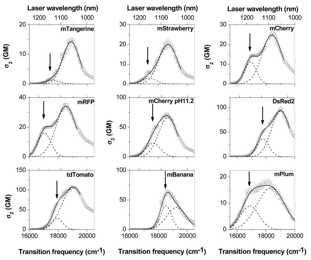
Intrinsically fluorescent proteins (FPs) exhibit broad variations of absorption and emission colors and are available for different imaging applications. The physical cause of the absorption wavelength change from 540 to 590 nm in the Fruits series of red FPs has been puzzling because the mutations that cause the shifts do not disturb the pi-conjugation pathway of the chromophore. Here, we use two-photon absorption measurements to show that the different colors can be explained by quadratic Stark effect due to variations of the strong electric field within the beta barrel. This model brings simplicity to a bewildering diversity of fluorescent protein properties, and it suggests a new way to sense electrical fields in biological systems.


Figures

Figure 1
Figure 1
Two-photon absorption spectra of a series of red FPs in the region of the first electronic transition. 2PA cross section σ2 (in GM, 1GM = 10−50 cm4 s) is plotted versus transition frequency (equal to twice the laser photon frequency). Laser wavelength used for excitation is shown as a top x-axis. The fit with a sum of two Gaussians is shown with continuous line. Individual Gaussians are shown by dashed lines. An arrow depicts the 0–0 1PA transition frequency which was kept fixed upon fitting of 2PA spectra.
Figure 2
Figure 2
Dependence of the pure electronic S0 → S1 transition frequency (which is very close to 1PA maximum) on permanent dipole moment difference between S1 and S0 states for a series of red FPs. Top x-axis shows the projection of effective electric field on the direction of Δμ 0. fS is the local field factor. Continuous line shows the best fit with second order polynomial: y = A + Bx + Cx 2 with the coefficients A = 19350 ± 116 cm−1, B = − 2180 ± 100 cm−1 D−1, C = 486 ± 20 cm−1 D−2. The inset shows the chromophore structure. Note that the crystal structures, available for DsRed (9), mStrawberry (10), mCherry (10), mPlum (11), and mBanana (12), show that the chromophore is the same for these five proteins and contains the acylimine group. For the remaining four proteins, there are strong indications that the chromophore structure is the same as shown. It is known that the originally produced acylimine tail of the DsRed-type chromophore can be attacked by either –OH group of Thr66 or –SH group of Cys66, to produce new chromophores in mOrange (10) and mKO (34), respectively. For this reaction to occur Glu215 should be deprotonated and, in the case of Cys66, position 70 should be occupied by Arg, not Lys. mRFP is a product of DsRed with no mutations in close proximity to the chromophore, including position 66. mRFP is also a progenitor for mCherry and mStrawberry, which both have the same chromophore as DsRed. tdTomato and mCherry (at pH11.2) have Q66M mutation, which should not have nucleophilic activity. mTangerine, similarly to mBanana holding intact acylimine tail, possesses Q66C mutation, but has Lys in position 70.

References

    1. Prasher DC, Eckenrode VK, Ward WW, Prendergast FG, Cormier MJ. Gene. 1992;111:229. - PubMed
    1. Chalfie M, Tu Y, Euskirchen G, Ward WW, Prasher DC. Science. 1994;263:802. - PubMed
    1. Tsien RY. Annu. Rev. Biochem. 1998;67:509. - PubMed
    1. Matz MV, Fradkov AF, Labas YA, Savitsky AP, Zaraisky AG, Markelov ML, Lukyanov SA. Nat. Biotech. 1999;17:969. - PubMed
    1. Shaner NC, Patterson GH, Davidson MW. J. Cell Sci. 2007;120:4247. - PubMed

Publication types

Substances