

High-resolution peripheral quantitative computed tomography can assess microstructural and mechanical properties of human distal tibial bone
- PMID: 19775199
- PMCID: PMC3130204
- DOI: 10.1359/jbmr.090822
High-resolution peripheral quantitative computed tomography can assess microstructural and mechanical properties of human distal tibial bone
Abstract
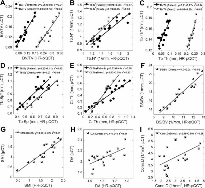
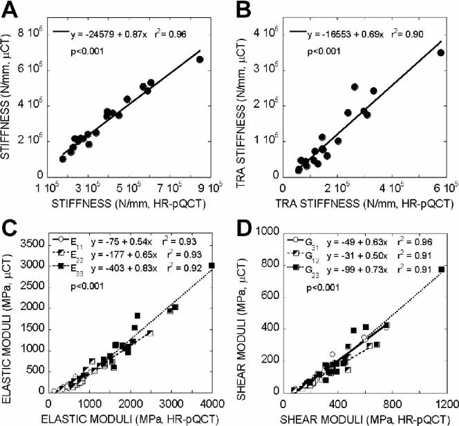
High-resolution peripheral quantitative computed tomography (HR-pQCT) is a newly developed in vivo clinical imaging modality. It can assess the 3D microstructure of cortical and trabecular bone at the distal radius and tibia and is suitable as an input for microstructural finite element (microFE) analysis to evaluate bone's mechanical competence. In order for microstructural and image-based microFE analyses to become standard clinical tools, validation with a current gold standard, namely, high-resolution micro-computed tomography (microCT), is required. Microstructural measurements of 19 human cadaveric distal tibiae were performed for the registered HR-pQCT and microCT images, respectively. Next, whole bone stiffness, trabecular bone stiffness, and elastic moduli of cubic subvolumes of trabecular bone in both HR-pQCT and microCT images were determined by microFE analysis. The standard HR-pQCT patient protocol measurements, derived bone volume fraction (BV/TV(d)), trabecular number (Tb.N*), trabecular thickness (Tb.Th), trabecular spacing (Tb.Sp), and cortical thickness (Ct.Th), as well as the voxel-based direct measurements, BV/TV, Tb.N*, Tb.Th*, Tb.Sp*, Ct.Th, bone surface-to-volume ratio (BS/BV), structure model index (SMI), and connectivity density (Conn.D), correlated well with their respective gold standards, and both contributed to microFE-predicted mechanical properties in either single or multiple linear regressions. The mechanical measurements, although overestimated by HR-pQCT, correlated highly with their gold standards. Moreover, elastic moduli of cubic subvolumes of trabecular bone predicted whole bone or trabecular bone stiffness in distal tibia. We conclude that microstructural measurements and mechanical parameters of distal tibia can be efficiently derived from HR-pQCT images and provide additional information regarding bone fragility.
Copyright 2010 American Society for Bone and Mineral Research.
Figures
Similar articles
-
Accuracy of high-resolution in vivo micro magnetic resonance imaging for measurements of microstructural and mechanical properties of human distal tibial bone.J Bone Miner Res. 2010 Sep;25(9):2039-50. doi: 10.1002/jbmr.92. J Bone Miner Res. 2010. PMID: 20499379 Free PMC article.
-
High-resolution peripheral quantitative computed tomography (HR-pQCT) can assess microstructural and biomechanical properties of both human distal radius and tibia: Ex vivo computational and experimental validations.Bone. 2016 May;86:58-67. doi: 10.1016/j.bone.2016.02.016. Epub 2016 Feb 26. Bone. 2016. PMID: 26924718
-
Validation of calcaneus trabecular microstructure measurements by HR-pQCT.Bone. 2018 Jan;106:69-77. doi: 10.1016/j.bone.2017.09.013. Epub 2017 Oct 3. Bone. 2018. PMID: 28986143
-
Fast trabecular bone strength predictions of HR-pQCT and individual trabeculae segmentation-based plate and rod finite element model discriminate postmenopausal vertebral fractures.J Bone Miner Res. 2013 Jul;28(7):1666-78. doi: 10.1002/jbmr.1919. J Bone Miner Res. 2013. PMID: 23456922 Free PMC article. Clinical Trial.
-
High-resolution imaging techniques for the assessment of osteoporosis.Radiol Clin North Am. 2010 May;48(3):601-21. doi: 10.1016/j.rcl.2010.02.015. Radiol Clin North Am. 2010. PMID: 20609895 Free PMC article. Review.
Cited by
-
A comparative study of trabecular bone micro-structural measurements using different CT modalities.Phys Med Biol. 2020 Nov 27;65(23):10.1088/1361-6560/abc367. doi: 10.1088/1361-6560/abc367. Phys Med Biol. 2020. PMID: 33086213 Free PMC article.
-
FOXO1 orchestrates the bone-suppressing function of gut-derived serotonin.J Clin Invest. 2012 Oct;122(10):3490-503. doi: 10.1172/JCI64906. Epub 2012 Sep 4. J Clin Invest. 2012. PMID: 22945629 Free PMC article.
-
High-resolution peripheral quantitative computed tomographic imaging of cortical and trabecular bone microarchitecture in patients with type 2 diabetes mellitus.J Clin Endocrinol Metab. 2010 Nov;95(11):5045-55. doi: 10.1210/jc.2010-0226. Epub 2010 Aug 18. J Clin Endocrinol Metab. 2010. PMID: 20719835 Free PMC article.
-
Accuracy of individual trabecula segmentation based plate and rod finite element models in idealized trabecular bone microstructure.J Biomech Eng. 2013 Apr;135(4):044502. doi: 10.1115/1.4023983. J Biomech Eng. 2013. PMID: 24231904 Free PMC article.
-
Skeletal structure in postmenopausal women with osteopenia and fractures is characterized by abnormal trabecular plates and cortical thinning.J Bone Miner Res. 2014;29(5):1101-9. doi: 10.1002/jbmr.2144. J Bone Miner Res. 2014. PMID: 24877245 Free PMC article. Clinical Trial.
References
-
- NIH Consensus Development Panel on Osteoporosis Prevention. Consensus Development Conference Report: Prophylaxis and treatment of osteoporosis. Osteoporosis Int. 1991;1:114–117. - PubMed
-
- Miller PD. Clinical use of bone mass measurements in adults for the assessment and management of osteoporosis. In: Favus MJ, editor. Primer on the Metabolic Bone Disease and Disorders of Mineral Metabolism. 6th ed. Washington, DC: American Society for Bone and Mineral Research; 2006. pp. 150–161.
-
- Sornay-Rendu E, Boutroy S, Munoz F, Delmas PD. Alterations of cortical and trabecular architecture are associated with fractures in postmenopausal women, partially independent of decreased BMD measured by DXA: the OFELY Study. J Bone Miner Res. 2007;22:425–433. - PubMed
-
- Goulet RW, Goldstein SA, Ciarelli MJ, Kuhn JL, Brown MB, Feldkamp LA. The relationship between the structural and orthogonal compressive properties of trabecular bone. J Biomech. 1994;27:375–389. - PubMed
-
- Kabel J, Odgaard A, van Rietbergen B, Huiskes R. Connectivity and the elastic properties of cancellous bone. Bone. 1999;24:115–120. - PubMed
