Review
. 2009 Oct;30(6):624-712.
doi: 10.1210/er.2009-0012. Epub 2009 Sep 23.

The mammalian ovary from genesis to revelation

Affiliations
Review

The mammalian ovary from genesis to revelation

Mark A Edson et al. Endocr Rev. 2009 Oct.

Abstract

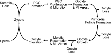
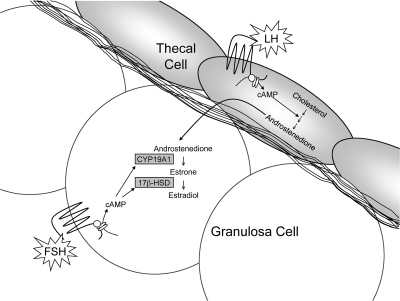
Two major functions of the mammalian ovary are the production of germ cells (oocytes), which allow continuation of the species, and the generation of bioactive molecules, primarily steroids (mainly estrogens and progestins) and peptide growth factors, which are critical for ovarian function, regulation of the hypothalamic-pituitary-ovarian axis, and development of secondary sex characteristics. The female germline is created during embryogenesis when the precursors of primordial germ cells differentiate from somatic lineages of the embryo and take a unique route to reach the urogenital ridge. This undifferentiated gonad will differentiate along a female pathway, and the newly formed oocytes will proliferate and subsequently enter meiosis. At this point, the oocyte has two alternative fates: die, a common destiny of millions of oocytes, or be fertilized, a fate of at most approximately 100 oocytes, depending on the species. At every step from germline development and ovary formation to oogenesis and ovarian development and differentiation, there are coordinated interactions of hundreds of proteins and small RNAs. These studies have helped reproductive biologists to understand not only the normal functioning of the ovary but also the pathophysiology and genetics of diseases such as infertility and ovarian cancer. Over the last two decades, parallel progress has been made in the assisted reproductive technology clinic including better hormonal preparations, prenatal genetic testing, and optimal oocyte and embryo analysis and cryopreservation. Clearly, we have learned much about the mammalian ovary and manipulating its most important cargo, the oocyte, since the birth of Louise Brown over 30 yr ago.


Figures

Figure 1
Figure 1
Reproductive life cycle of a mammalian female. An oocyte and a spermatozoon will fuse to form a zygote and undergo multiple steps in embryogenesis. At about E6.5 in mouse, the PGC will be allocated and go through multiple steps to reach the genital ridge. In an XX mammal, the germ cell will form an oocyte that arrests at meiosis I (MI). During prenatal life in women, and in the perinatal period in mice, the oocyte will be encased in somatic cells to become primordial follicles. Upon recruitment into the growing pool, the oocyte increases in size during folliculogenesis. The LH surge will induce resumption of meiosis, release of the first body, arrest at MII, and subsequent ovulation of the oocyte into the fallopian tube. Fertilization with a spermatozoon will induce the completion of meiosis and release of the second polar body. The cycle continues in the next generation of females. During the reproductive cycle, there are multiple steps where significant oocyte loss is observed.
Figure 2
Figure 2
Gonad function and sexual differentiation. As shown, several major gene products influence the formation of the bipotential gonad (light gray), the development of the ovary (white), and the development of the testis (dark gray). SF1 is a central player in the bipotential gonad, being regulated by WT1, LHX9, and M33 (CBX2), and at other steps in gonadal differentiation. In XY gonads, SRY functions in a short window in pre-Sertoli cells to up-regulate the transcription of SOX9 that is already expressed at low levels through the action of SF1. This higher SOX9 expression then suppresses SRY in a negative feedback loop and also up-regulates itself through the combined actions of SF1 and SOX9 on the SOX9 promoter. SOX9 also up-regulates FGF9 that signals back through FGFR2 to maintain/increase SOX9 expression. The ovarian differentiation pathway involves RSPO1 increasing the signaling of WNT4, which up-regulates β-catenin. β-catenin acts to up-regulate WNT4 and other proteins such as FST. The testis pathway appears to mainly antagonize this pathway through decreasing β-catenin levels. Likewise, β-catenin antagonizes the testis pathway by destabilizing SOX9.
Figure 3
Figure 3
Sexually dimorphic initiation of meiosis in the embryonic ovary. During embryogenesis, the mesonephroi adjacent to the developing ovary (A) and testes (B) contain several aldehyde dehydrogenases that convert retinaldehyde to all-trans-retinoic acid (RA). The somatic cells of the developing testes contain the enzyme CYP26B1, which degrades RA to pass freely to the germ cell to bind to retinoic acid receptors (RAR). In the developing ovary, RA induces STRA8, which induces SYCP3, which is stably translated in the presence of DAZL and becomes chromosomally localized as the XX germ cell becomes an oocyte and enters meiosis. In male germ cells, the absence of high enough levels of RA early and under the repressive actions of NANOS at later time points, STRA8 is not synthesized and the XY germ cell becomes mitotically arrested.
Figure 4
Figure 4
Classification of the major stages of mammalian folliculogenesis. Primordial follicles form 1–2 d after birth in mice and in utero in humans. Preantral follicles begin to develop prenatally in humans, whereas in mice this occurs postnatally. In both mice and humans, preantral follicular development does not require stimulation by the pituitary gonadotropins. By the secondary stage, an additional layer of somatic cells, the theca, forms outside the basement membrane of the follicle. At puberty, FSH secreted by the pituitary promotes further granulosa cell proliferation and survival. Ovulation of the dominant follicle occurs in response to a rise in the other pituitary gonadotropin, LH. After ovulation, the remaining granulosa and theca cells undergo terminal differentiation to form the CL. In most cases, primordial, primary, secondary, preantral, and antral are names commonly used to refer to the different stages of folliculogenesis; however, a classification system described by Pedersen and Peters (768) is also used. Pedersen stages are determined based on the size of the oocyte and the number of granulosa cells in cross-section for any given follicle. Although not shown, certain Pedersen stages are subdivided (i.e., 3a, 3b, 5a, 5b) depending on the number of granulosa cells surrounding the oocyte.
Figure 5
Figure 5
The two-cell, two-gonadotropin concept of follicular steroid production. The main function of thecal cells during folliculogenesis is the production of steroids. Although thecal cells are capable of de novo production of androgens, they lack aromatase (CYP19A1), which is required to convert androgens into estradiol. Thecal cells respond to basal levels of LH by up-regulating biosynthetic enzymes involved in steroid production, including STAR, CYP11A1, CYP17A1, and 3β-hydroxysteroid dehydrogenase (3β-HSD). STAR facilitates the transport of cholesterol to the inner mitochondrial membrane, where it is converted to pregnenolone by CYP11A1. Pregnenolone is converted to dehydroepiandrosterone (DHEA) by CYP17A1. Finally, 3β-HSD converts DHEA into androstenedione, which diffuses across the basement membrane to granulosa cells. In response to stimulation by FSH, granulosa cells up-regulate CYP19A1 and 17β-hydroxysteroid dehydrogenase (17β-HSD), which convert androstenedione into estradiol (249).
Figure 6
Figure 6
Hyaluronan-IαI-PTX3 interactions stabilize the cumulus matrix. GDF9 and BMP15 secreted by the oocyte stimulate cumulus cells to produce HAS2, TNFAIP6, and PTX3. HAS2 catalyzes the synthesis of hyaluronan (HA; curved line), the structural backbone of the cumulus matrix. Hyaluronan is covalently linked to the heavy chain of IαI (white box) by the catalytic activity of TNFAIP6. Multimers of PTX3 (black trapezoids) stabilize the hyaluronan matrix by interacting with IαI.
Figure 7
Figure 7
Dicer-dependent synthesis of miRNAs and endogenous siRNAs. A, miRNA genes are transcribed by RNA polymerase II to generate primary transcripts. These transcripts are cropped by the DROSHA/DGCR8 Microprocessor complex to yield approximately 70-nt precursor miRNAs, which are exported from the nucleus to the cytoplasm by Exportin 5. There, the DICER complex processes the precursor into a 21- to 23-nt duplex consisting of the mature miRNA and its antisense. The mature miRNA is preferentially loaded into the miRNA-induced silencing complex (miRISC), which mediates pairing with complementary sequences in the 3′ UTR of target mRNA transcripts, leading to mRNA degradation and translational repression. The specificity of targeting is especially dependent on nucleotides 2–8 of the mature miRNA, known as the seed sequence. B, Endogenous siRNAs are synthesized from long, double-stranded RNA precursors derived from repetitive sequences, sense-antisense pairs, or inverted repeats that form hairpins. It is unknown whether endogenous siRNA precursors use Exportin 5 to translocate to the nucleus; however, once in the cytoplasm they are processed by the DICER complex into 21- to 23-nt duplexes. Mature siRNAs are incorporated into the siRNA-induced silencing complex (siRISC), which is similar to the miRISC but may also have unique components. Endogenous siRNAs function in transposon suppression and other functions such as pseudogene regulation of founding source mRNAs.

References

    1. Chiquoine AD 1954 The identification, origin and migration of the primordial germ cells in the mouse embryo. Anat Rec 118:135–146 - PubMed
    1. Mintz B, Russell ES 1957 Gene-induced embryological modifications of primordial germ cells in the mouse. J Exp Zool 134:207–237 - PubMed
    1. McCoshen JA, McCallion DJ 1975 A study of the primordial germ cells during their migratory phase in Steel mutant mice. Experientia 31:589–590 - PubMed
    1. Ozdzenski W 1967 Observations on the origin of the primordial germ cells in the mouse. Zool Polon 17:367–379
    1. Spiegelman M, Bennett D 1973 A light- and electron-microscopic study of primordial germ cells in the early mouse embryo. J Embryol Exp Morphol 30:97–118 - PubMed

Publication types