. 2009 Sep 23;29(38):11982-92.
doi: 10.1523/JNEUROSCI.3158-09.2009.

CD14 and toll-like receptors 2 and 4 are required for fibrillar A{beta}-stimulated microglial activation

Affiliations

CD14 and toll-like receptors 2 and 4 are required for fibrillar A{beta}-stimulated microglial activation

Erin G Reed-Geaghan et al. J Neurosci. .

Abstract

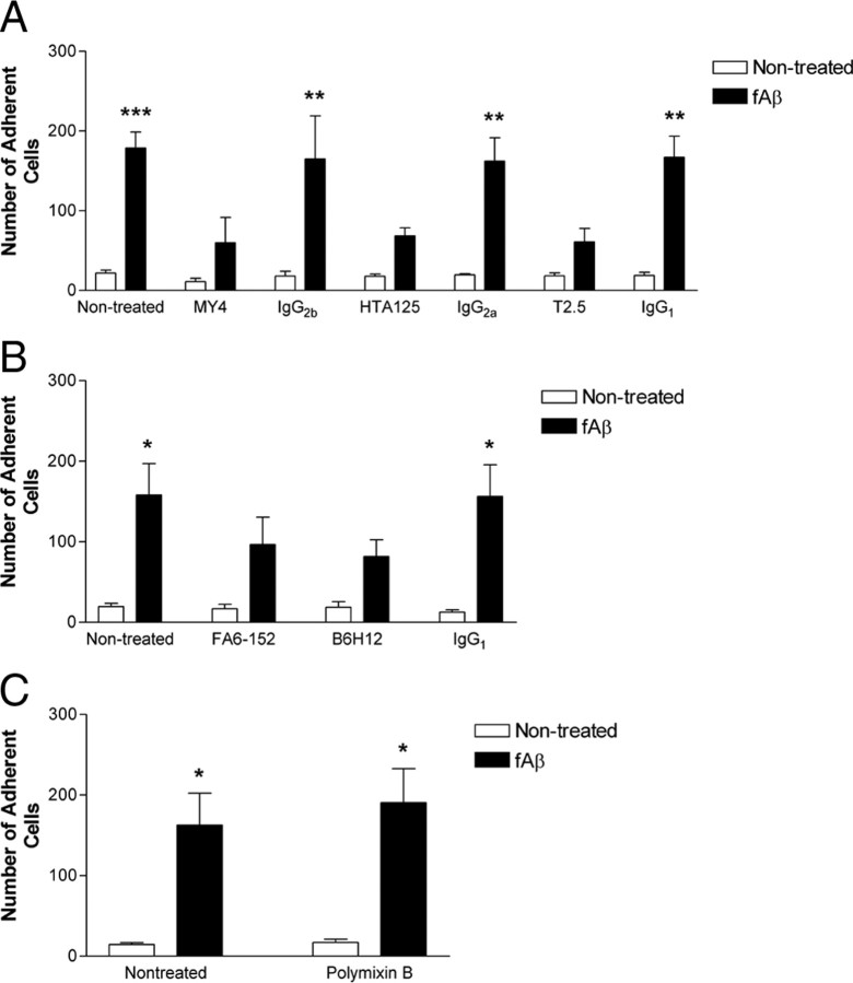
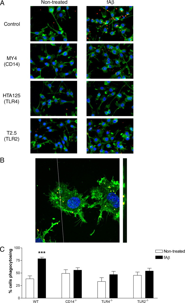
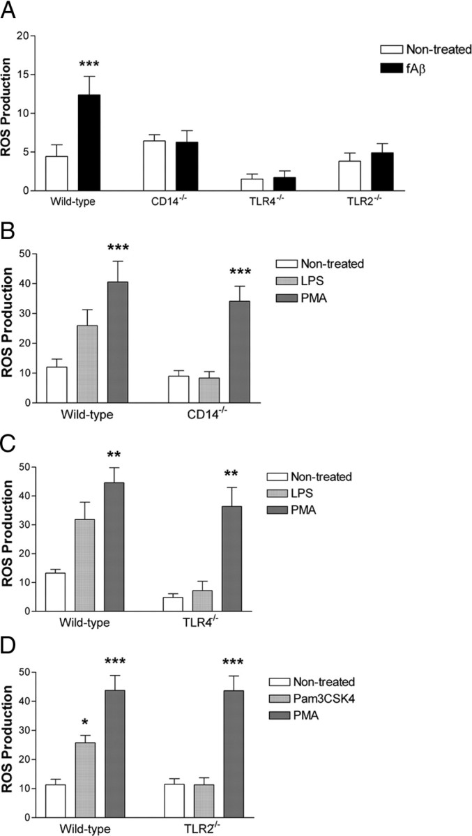
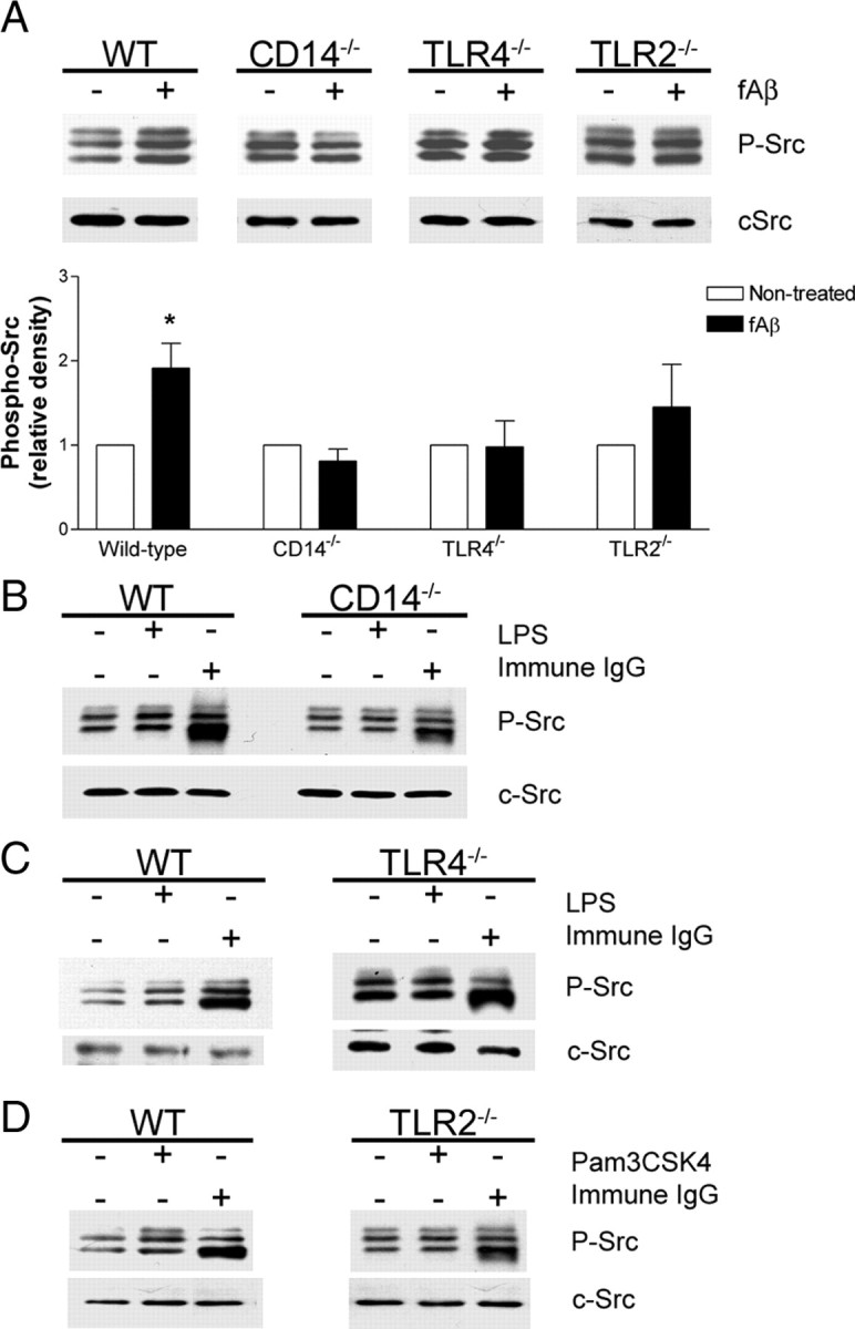
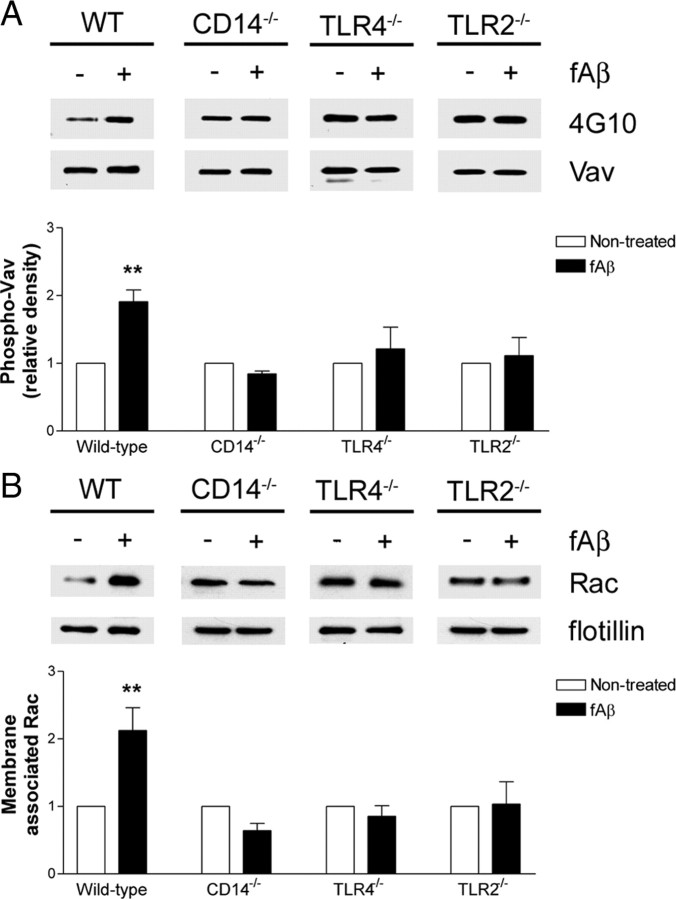
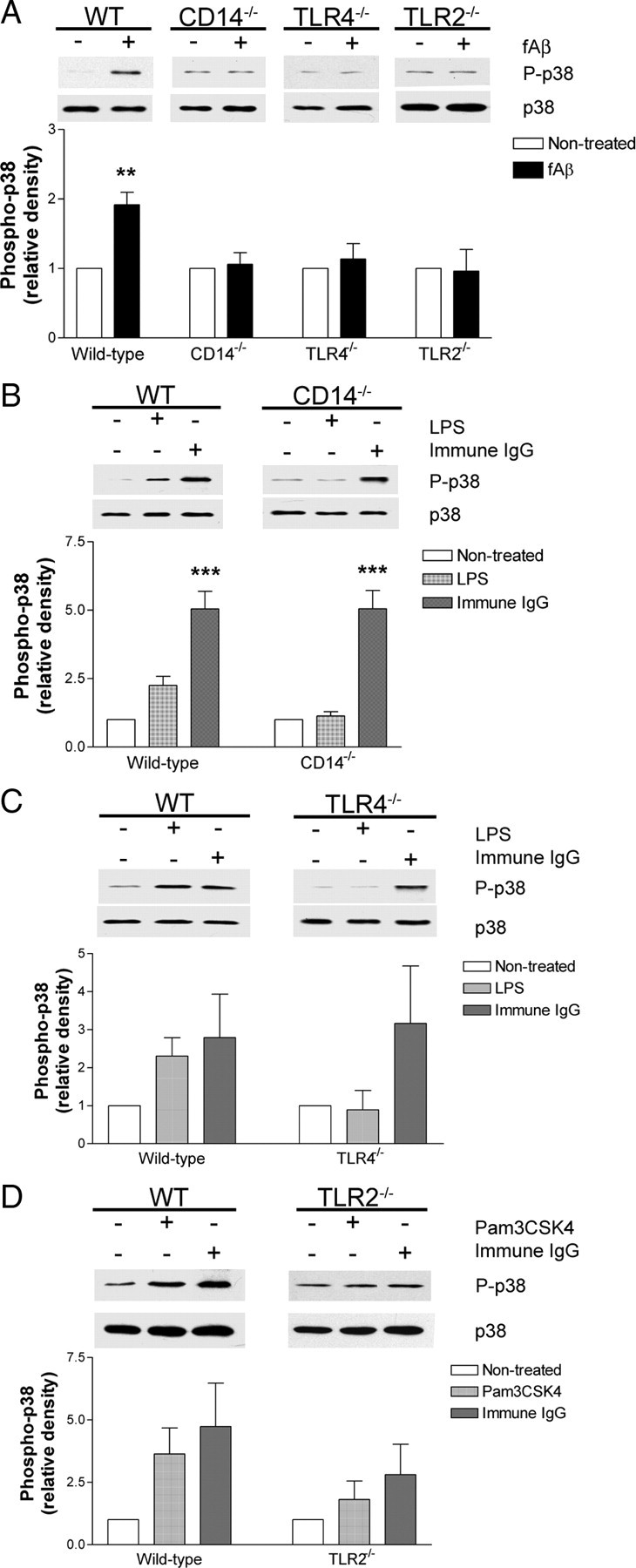
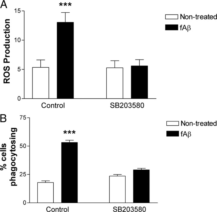
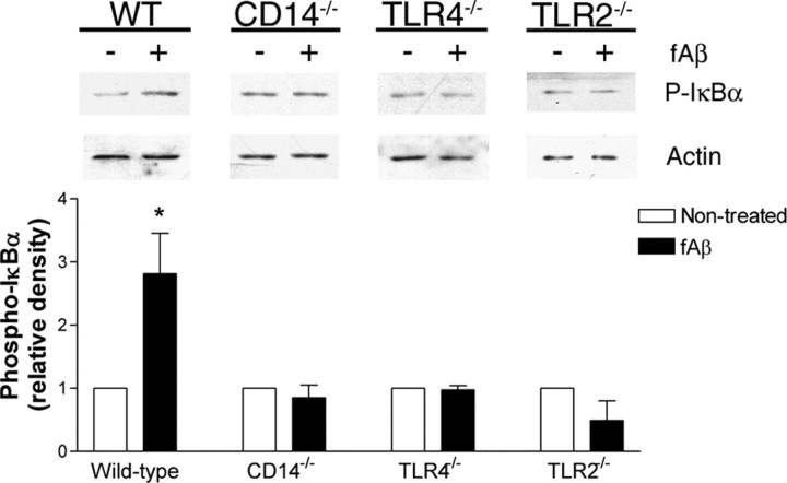
Microglia are the brain's tissue macrophages and are found in an activated state surrounding beta-amyloid plaques in the Alzheimer's disease brain. Microglia interact with fibrillar beta-amyloid (fAbeta) through an ensemble of surface receptors composed of the alpha(6)beta(1) integrin, CD36, CD47, and the class A scavenger receptor. These receptors act in concert to initiate intracellular signaling cascades and phenotypic activation of these cells. However, it is unclear how engagement of this receptor complex is linked to the induction of an activated microglial phenotype. We report that the response of microglial cells to fibrillar forms of Abeta requires the participation of Toll-like receptors (TLRs) and the coreceptor CD14. The response of microglia to fAbeta is reliant upon CD14, which act together with TLR4 and TLR2 to bind fAbeta and to activate intracellular signaling. We find that cells lacking these receptors could not initiate a Src-Vav-Rac signaling cascade leading to reactive oxygen species production and phagocytosis. The fAbeta-mediated activation of p38 MAPK also required CD14, TLR4, and TLR2. Inhibition of p38 abrogated fAbeta-induced reactive oxygen species production and attenuated the induction of phagocytosis. Microglia lacking CD14, TLR4, and TLR2 showed no induction of phosphorylated IkappaBalpha following fAbeta. These data indicate these innate immune receptors function as members of the microglial fAbeta receptor complex and identify the signaling mechanisms whereby they contribute to microglial activation.


Figures

Figure 1.
Figure 1.
CD14, TLR4, and TLR2 mediate the recognition and binding of fAβ. A, B, THP-1 monocytes were incubated in the presence or absence of function blocking antibodies to CD14 (MY4, A), TLR4 (HTA125, A), TLR2 (T2.5, A), CD36 (FA6-152, B), CD47 (B6H12, B), or their isotype controls (IgG2b, IgG2a, IgG1). Cells were then added to fAβ bound to a glass slide and the number of adherent cells was determined. C, THP-1 monocytes were incubated in the presence or absence of 0.1 μg/ml polymixin B. Cells were then added to fAβ bound to a glass slide and the number of adherent cells was determined. The data shown are the mean ± SEM of three independent experiments. *p < 0.05, **p < 0.01, ***p < 0.001 compared with Non-treated.
Figure 2.
Figure 2.
Phagocytosis stimulated by fAβ requires CD14, TLR4, and TLR2. A, BV2 microglia were pretreated with function blocking antibodies to CD14 (MY4), TLR4 (HTA125), or TLR2 (T2.5), before stimulation with fAβ. Fluorescent microspheres were then added for 30 min. Cells were stained with phalloidin for visualization of actin and DAPI. Magnification is 40×. B, Confocal images (100×) of BV2 microglia treated with fAβ-containing microspheres in both the x–y plane (left) and z-scan cut (right) through the cells. C, Primary microglia from C57BL/6 (WT), CD14−/−, TLR4−/−, or TLR2−/− mice were stimulated with fAβ, followed by incubation with fluorescent microspheres. The fraction of cells containing microspheres was determined by counting three random fields (>100 cells) on an inverted microscope. The data shown are the mean ± SEM of three independent experiments performed in duplicate. ***p < 0.001 compared with Non-treated.
Figure 3.
Figure 3.
CD14, TLR4, and TLR2 are necessary for fAβ-stimulated reactive oxygen species production. A–D, Primary microglia from C57BL/6 (WT), CD14−/−, TLR4−/−, TLR2−/− mice were stimulated with fAβ (A), LPS (B, C), Pam3CSK4 (D), or PMA (B–D). Reduction of NBT results in the formation of a dark blue formazan deposit viewable by a light microscope. Three random fields (>100 cells) were counted and the fraction of cells with blue formazan deposits was determined. The data shown are the mean ± SEM of three independent experiments performed in duplicate. *p < 0.05, **p < 0.01, ***p < 0.001 compared with Non-treated.
Figure 4.
Figure 4.
Activation of the Src tyrosine kinases by fAβ requires CD14, TLR4, and TLR2. A–D, Primary microglia from C57BL/6 (WT), CD14−/−, TLR4−/−, or TLR2−/− mice were stimulated with fAβ (A), LPS (B, C), Pam3CSK4 (D), or immune IgG (B–D). Cell lysates were analyzed by Western blot analysis using an anti-phospho-Src family antibody. Blots were stripped and reprobed with an anti-cSrc antibody as a protein loading control. A, Western blot band intensity of phosphorylated Src kinases was normalized to cSrc levels and expressed as relative density. Western blots are representative, and the densitometry data are the mean ± SEM from at least three independent experiments. *p < 0.05 compared with Non-treated.
Figure 5.
Figure 5.
CD14, TLR4, and TLR2 are required for fAβ-induced phosphorylation of the Rac GEF Vav and subsequent translocation of Rac to the membrane. A, Primary microglia from C57BL/6 (WT), CD14−/−, TLR4−/−, or TLR2−/− mice were stimulated with fAβ. Vav was immunoprecipitated from cell lysates with an anti-Vav antibody and analyzed by Western blot analysis using the anti-phosphoTyr antibody 4G10. Blots were stripped and reprobed with an anti-Vav antibody as a protein loading control. B, Primary microglia from C57BL/6 (WT), CD14−/−, TLR4−/−, or TLR2−/− mice were stimulated with fAβ. Membrane fractions were analyzed by Western blot analysis with an anti-Rac antibody and an anti-flotillin antibody as a protein loading control. Western blots are representative, and the densitometry data are the mean ± SEM from three independent experiments. **p < 0.01 compared with Non-treated.
Figure 6.
Figure 6.
Activation of the p38 MAP kinase by fAβ requires CD14, TLR4, and TLR2. A–D, Primary microglia from C57BL/6 (WT), CD14−/−, TLR4−/−, or TLR2−/− mice were stimulated with fAβ (A), LPS (B, C), Pam3CSK4 (D), or immune IgG (B–D). Cell lysates were analyzed by Western blot analysis using an anti-phospho-p38 antibody. Blots were stripped and reprobed with an anti-p38 antibody as a protein loading control. Western blot band intensity of phosphorylated-p38 was normalized to p38 levels and expressed as relative density. Western blots are representative, and the densitometry data are the mean ± SEM from at least three independent experiments. **p < 0.01, ***p < 0.001 compared with Non-treated.
Figure 7.
Figure 7.
Fibrillar Aβ-stimulated ROS production and phagocytosis requires the activity of p38. A, Primary microglia from C57BL/6 mice were pretreated with SB203580 before stimulation with fAβ. Reduction of NBT results in the formation of a dark blue formazan deposit viewable by a light microscope. Three random fields (>100 cells) were counted and the fraction of cells with blue formazan deposits was determined. The data shown are the mean ± SEM of three independent experiments performed in duplicate. ***p < 0.001 compared with Non-treated. B, BV2 microglia were pretreated with SB203580 before stimulation with fAβ. Fluorescent microspheres were added, following which the fraction of cells containing microspheres was determined by counting three random fields (>100 cells) on an inverted microscope. The data shown are the mean ± SEM of three independent experiments performed in duplicate. ***p < 0.001 compared with Non-treated.
Figure 8.
Figure 8.
CD14, TLR4, and TLR2 are required for fAβ-induced NFκB activation. Primary microglia from C57BL/6 (WT), CD14−/−, TLR4−/−, or TLR2−/− mice were stimulated with fAβ. Cell lysates were analyzed by Western blot analysis using an anti-phospho-IκBα antibody. Blots were stripped and reprobed with an anti-actin antibody as a protein loading control. Western blot band intensity of phosphorylated-IκBα was normalized to actin levels and expressed as relative density. Western blots are representative, and the densitometry data represent the mean ± SEM from at least three independent experiments. *p < 0.05 compared with Non-treated.

References

    1. Aderem A. How to eat something bigger than your head. Cell. 2002;110:5–8. - PubMed
    1. Aderem A, Underhill DM. Mechanisms of phagocytosis in macrophages. Annu Rev Immunol. 1999;17:593–623. - PubMed
    1. Ago T, Nunoi H, Ito T, Sumimoto H. Mechanism for phosphorylation-induced activation of the phagocyte NADPH oxidase protein p47(phox). Triple replacement of serines 303, 304, and 328 with aspartates disrupts the SH3 domain-mediated intramolecular interaction in p47(phox), thereby activating the oxidase. J Biol Chem. 1999;274:33644–33653. - PubMed
    1. Ago T, Kuribayashi F, Hiroaki H, Takeya R, Ito T, Kohda D, Sumimoto H. Phosphorylation of p47phox directs phox homology domain from SH3 domain toward phosphoinositides, leading to phagocyte NADPH oxidase activation. Proc Natl Acad Sci U S A. 2003;100:4474–4479. - PMC - PubMed
    1. Akira S. Toll-like receptors and innate immunity. Adv Immunol. 2001;78:1–56. - PubMed

Publication types

MeSH terms